 ENG
ENG


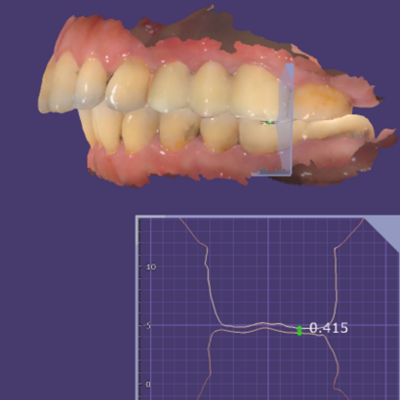
Table of Contents
The intraoral scanner is now becoming a popular equipment in the dental industry. Dentists usually use it to capture the intraoral data to replace the physical impression which may cause patients vomit or nausea. Besides, the intraoral scanner is usually cost and time-saving by getting rid of several traditional steps, such as transporting impressions, making models, and scanning physical models. With such advantages, it is widely accepted that more and more dentists turn to intraoral scanners and start up their first step of digital diagnostic and treatment, especially for occlusion data.
However, even though the intraoral scanner has been widely used, some users are always wondering how to make sure the occlusion data we acquired is accurate and the same with that in the patient’s mouth. This article will talk about this point and discuss it in detail.
What is occlusion?
Occlusion, technologically, is a relationship between the maxillary and mandibular teeth when they approach each other, during chewing or at rest. It is very important and plays a critical role in implantology, orthodontics and prosthodontics. To capture an accurate occlusion by intraoral scanner, there are lots of things we need to consider.
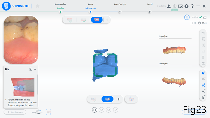
Common Problems of Occlusion Data During Intraoral Scan
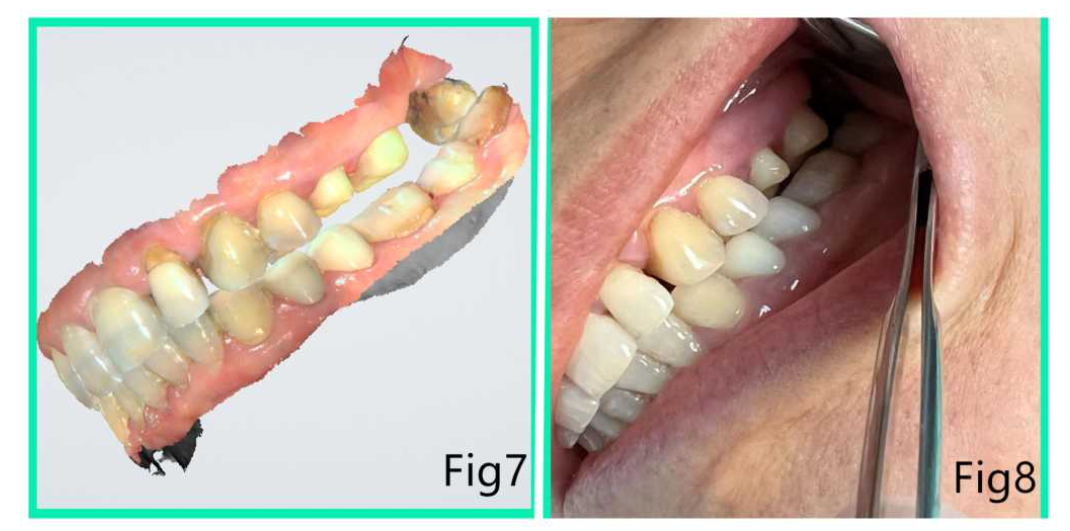
Teeth Data Staggered
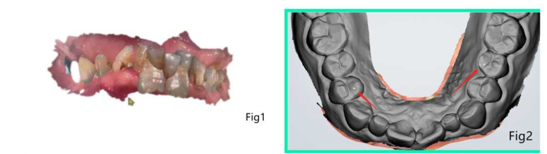
The Data of the Upper and Lower Gingiva are Staggered
A Deranged, Elevated Occlusion
Above are three common problems during occlusion scanning. What are the problems with the confused occlusal?
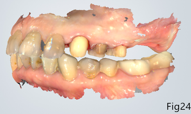
The Problem Results from Incorrect Occlusion
First, the incorrect occlusion leads to increasing difficulty in designing and manufacturing.
For lab technicians, if there is no intra-oral photograph or no obvious occlusion wear, it is hard to make a judgement whether the scan data is consistent with that in the mouth. If rescan is not possible in reality as the patients may already leave clinics, the most common method is to print out the model and reconstruct the maxillary frame to determine the correct bite.

The technician was unable to determine the value of the empty occlusion when all sides, including the left side, front side and right side, are empty. Without the accurate value of the empty occlusion, there is no way to make an accurate restoration.
Second, improper prostheses lead to a poor experience during dental treatment.
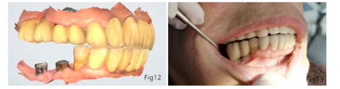
The inaccurate occlusion results in the emptiness or elevation of the final restoration, which leads to long-time occlusion adjustment, bringing bad dental experience to patients.
Factors Contributed to the Final Restoration’s Misfitted Occlusion: Clinic-side
- The occlusal relationship was not accurately estimated before the intraoral scan
Suggestion from SHINING 3D:
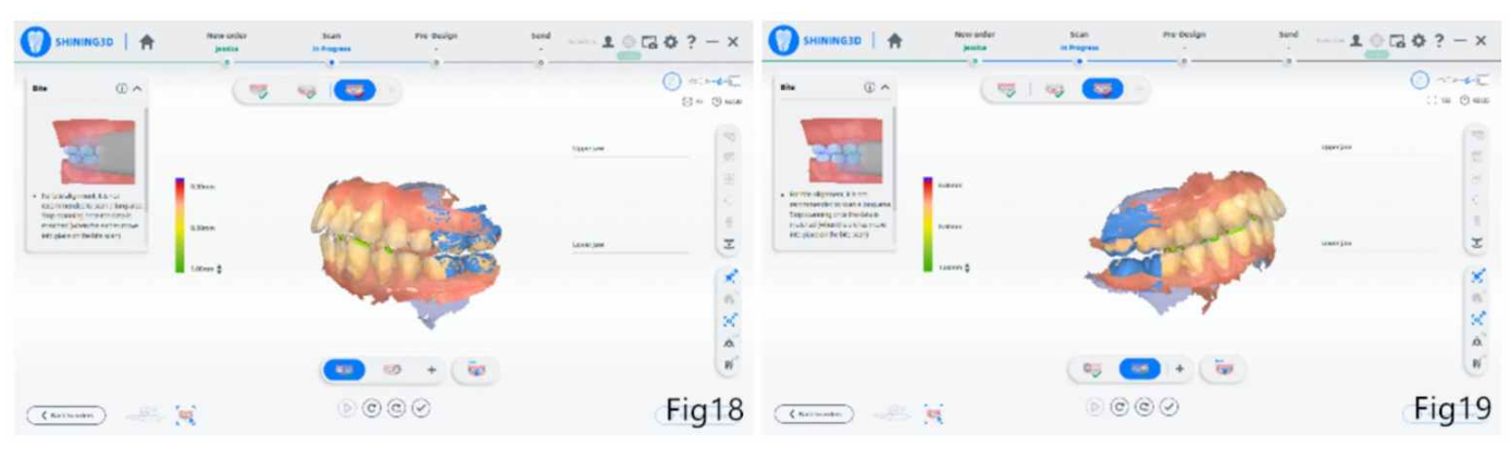
When scanning occlusal, it is necessary to confirm the accurate occlusal relationship first, then click the start button to scan.
During the scan process, we can hold the patient’s jaw by hand to ensure that the patient’s occlusion is fixed and does not move.
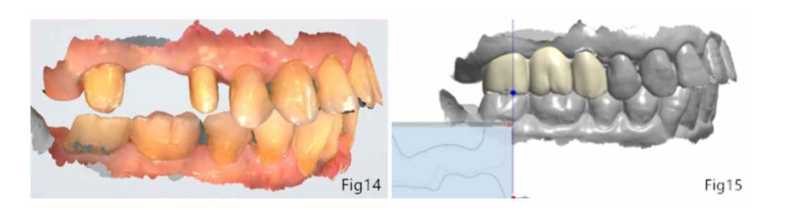
2. The scan range of unilateral occlusion is too large
Suggestion from SHINING 3D:
Selection of occlusal range: If the patient is scanned for full-mouth data, we recommend that the occlusal range of the left and right sides be captured, with 2~3 teeth on each side. The occlusal accuracy of the region with teeth in is higher than that in the edentulous region.
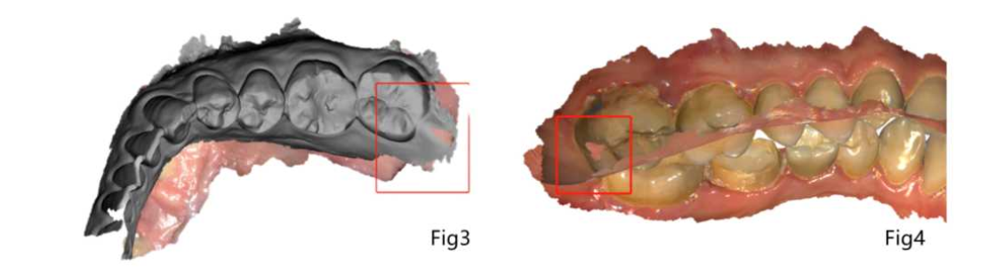
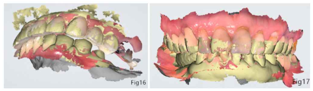
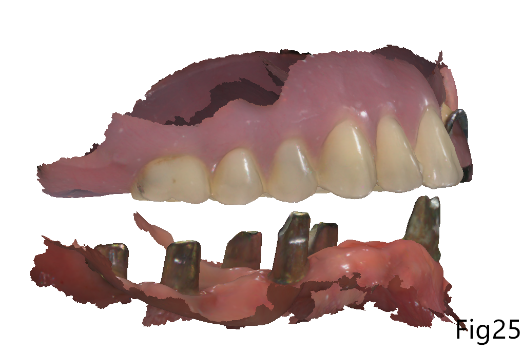
3. Excess soft tissue data in the working model
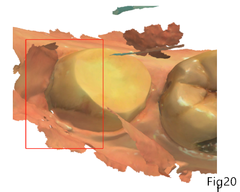
Suggestion from SHINING 3D:
It is recommended to capture the gingival data 4mm below the neck margin line, and active AI intelligent scanning function throughout the whole process.
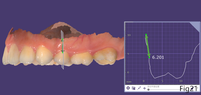
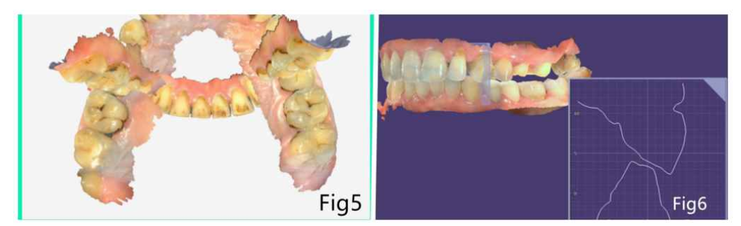
4. There is excess soft tissue data when occlusion aligns
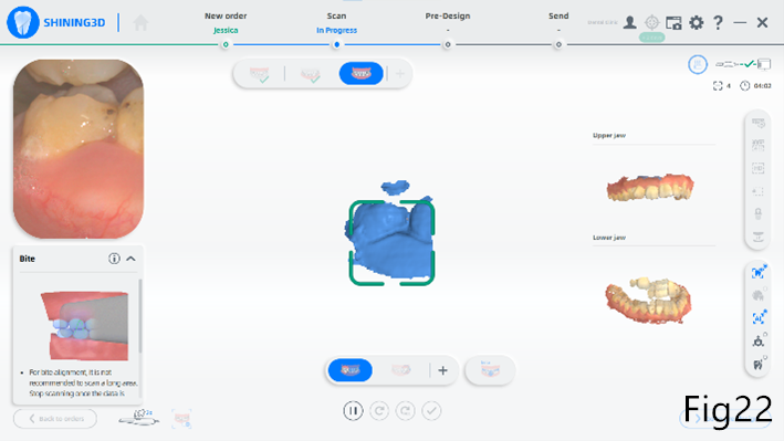
Suggestion from SHINING 3D:
Please remove the excess data when scanning occlusion, and make sure there is no interference during this process, otherwise, it will affect the occlusion accuracy.
Factors Contributed to the Final Restoration’s Misfitted Occlusion: Lab-side
- There are errors during the occlusion data transmission from the clinic to the lab
Suggestion from SHINING 3D:
It is recommended for some complex cases, such as orthodontics treatment, full mouth occlusion reconstruction, and free end restorations, if possible, it is much better to provide the bite wax to help technicians judge the patient’s occlusion more accurately.
2. The deviation in design and manufacture, especially in cases with unstable occlusion
Suggestion from SHINING 3D:
For cases like orthodontics treatment, full mouth occlusion reconstruction, and free end cases, the occlusion is always not stable, it is recommended that technicians print out the model and use bite wax to reconstruct the occlusion with the articulator in a traditional way.
In this article, we make an analysis of the reasons which may cause inaccurate occlusion from the clinic side and lab side, and the problems that result from incorrect occlusion, meanwhile, there are some tips to avoid these problems. Hope it can help you to get an accurate occlusion.