 ENG
ENG


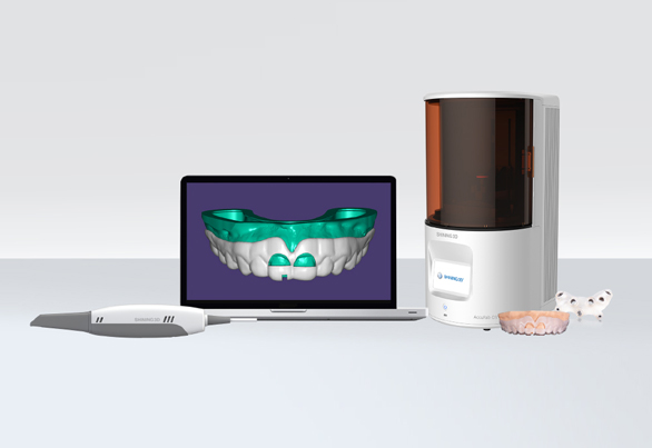
Table of Contents
Purpose
- Expose more healthy tooth tissue in accordance with the principle of periodontal biological width;
- Remove certain gingival and alveolar bone by surgical method;
- Increase the exposure of teeth, so as to carry out further repair or improve the aesthetic appearance of gingival shape.
The intention of crown lengthening is to make the gingival margin reach the predetermined position as accurately as possible. In the process of making treatment plan, the two basic principles to be considered in the surgical design are biological width and keratinized gingival width. The amount of gingiva can be accurately retained if there is a guide for crown lengthening surgery.
Products required
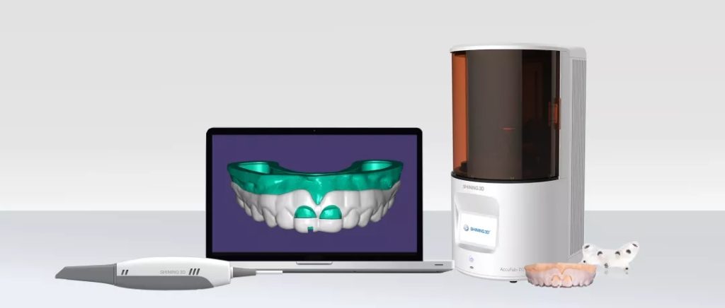
- Intraoral scanner: Aoralscan
- CAD software: Exocad
- 3D printer: AccuFab-D1
Workflow To Create A Surgical Guide for Crown Lengthening
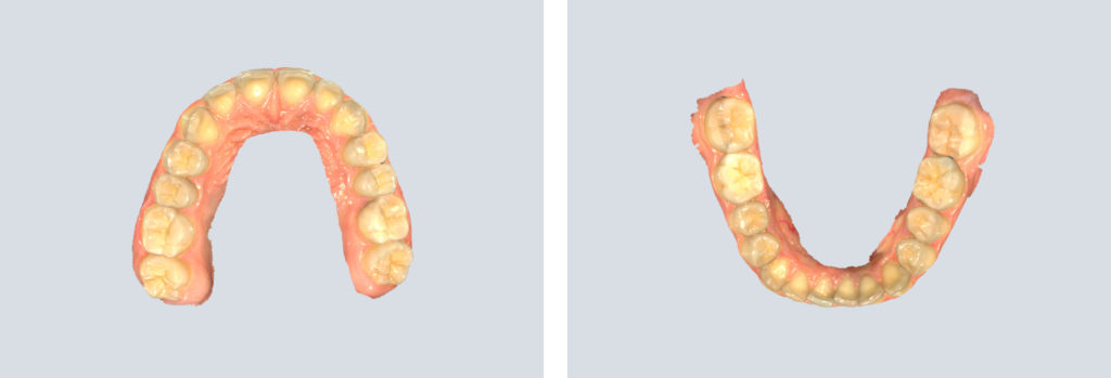
1. Data acquisition
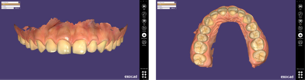
Use Aoralscan to capture as much mucous membrane data as possible to ensure sufficient design range for the guide in the following steps.
2. Exocad
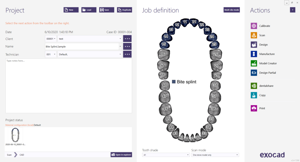
2.1 Create order
Create new order in Exocad.
2.2 Load data scanned by Aoralscan
Import data that gingival surgery requires.
3.3 Design bottom of the guide
You can set parameters and undercut of the guide in this step.
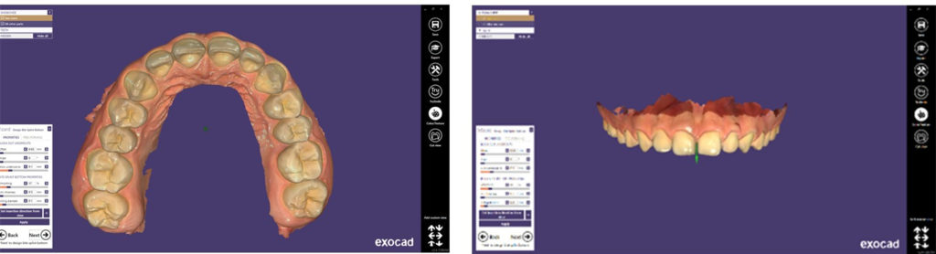
Note: Don’t block out the area where the gingiva will be treated.
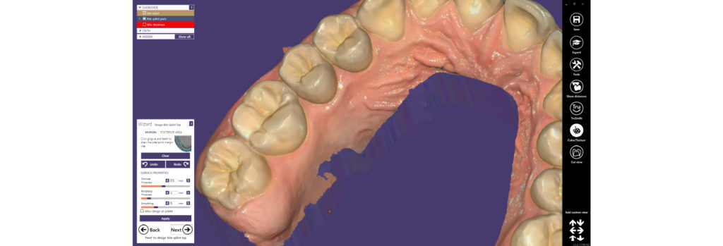
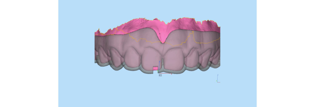
2.4 Draw margin line
Draw the margin line of the guide. Note that the margin line should not compress the frenum, etc., or else it will affect the fitting of the guide.
2.5 Generate guide
Generate guide and check if the margin line is in place. Adjust it if necessary.
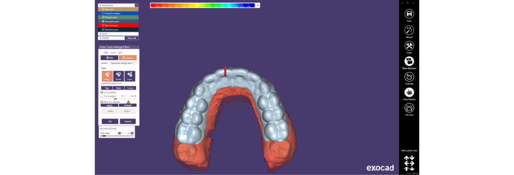
2.6 Free-form the guide
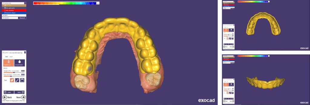
2.7 Design observation hole
2.8 Trim the gingiva area
Reference is obtained by setting the transparency of the guide. The area that needs to be extended of the guide can be cut off.
3. Print model & guide with AccuFab-D1
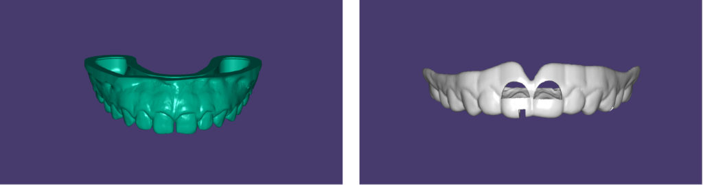
The finished prints:
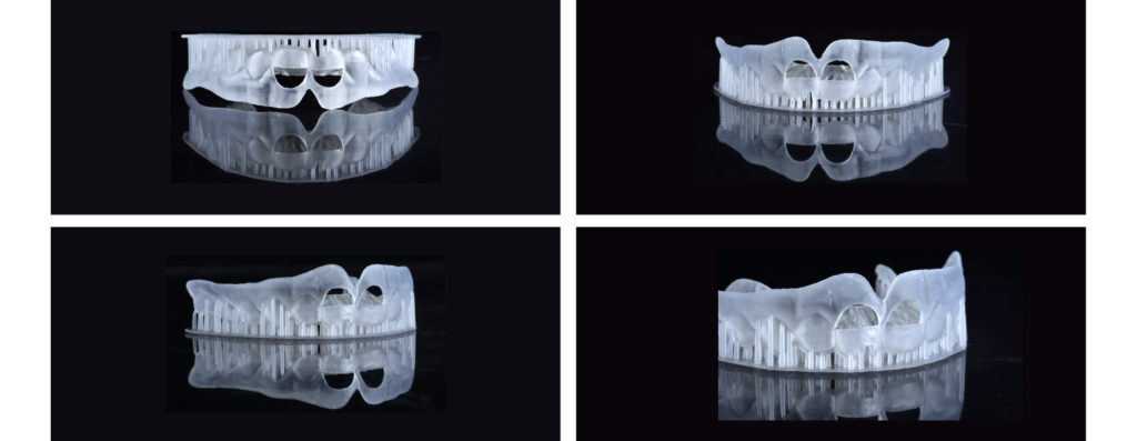
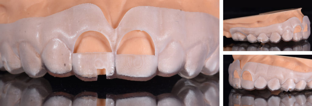
4. Try-in on the printed surgical guide for crown lengthening
The key to the success of crown lengthening
- Strictly master the indications of crown lengthening.
- Complete preoperative evaluation and detailed preoperative examination.
- Restoration-oriented design.
- Use of diagnostic models and diagnostic temporary restoration
- Surgical selection (based on the width of keratinized gingival and the distance between alveolar crest and CEJ)
- Intraoperative reconstructed biological width (at least 3 mm from the edge of the future prosthesis to the alveolar crest)
- Timing of postoperative permanent restoration (3~6 months).