 ENG
ENG


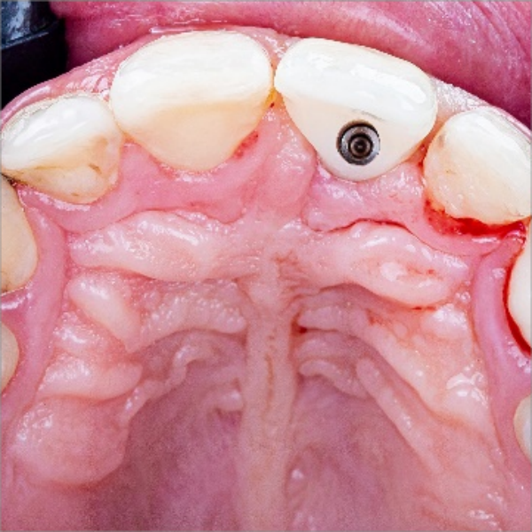
Table of Contents
As the world becomes increasingly digital, it’s no surprise that even the dental industry is embracing technology to make their daily work easier and more effective. Intraoral scanners and 3D printers are becoming more common in dental practices, allowing dentists to create precise restorations with greater efficiency than ever before. In a recent case study, provided by Dr. Abdelrahman Khalaf and Dr. Kirollos Hany of the Faculty of Dentistry at Assiut University in Egypt, they demonstrated how this technology can be used to restore anterior aesthetics zone – an area where appearance is particularly important.
Case Profile
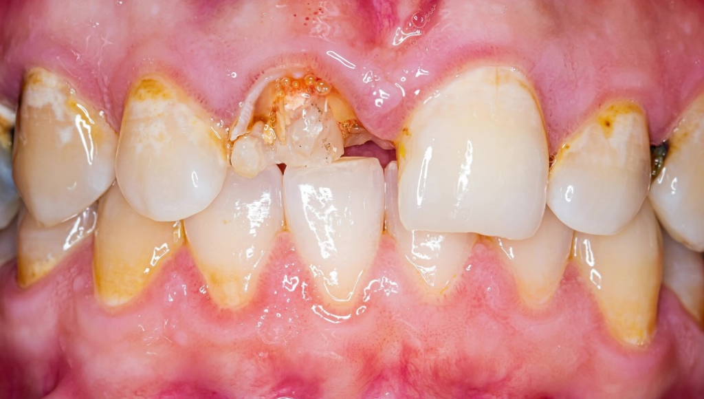
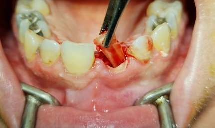
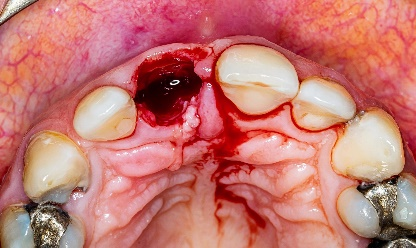
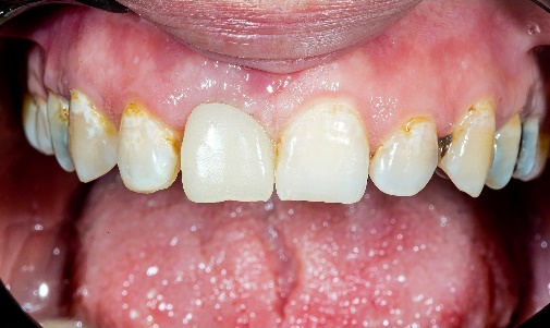
The female patient had been experiencing a damaged anterior tooth #11 for some time and sought treatment to restore both her chewing function and beautiful smile.
Make Treatment Plan
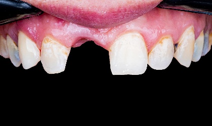
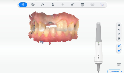
Upon her arrival, the dentist utilized Aoralscan 3 to scan her intraoral data and formulate a treatment plan that involved extracting tooth #11 and performing an implant surgery to support zirconia restorations. The comprehensive decision-making process took into account factors such as cost, treatment duration, and anticipated outcomes.
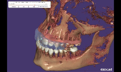
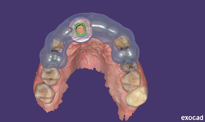
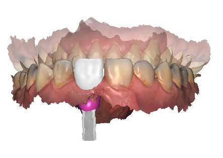
Make Surgical Guide
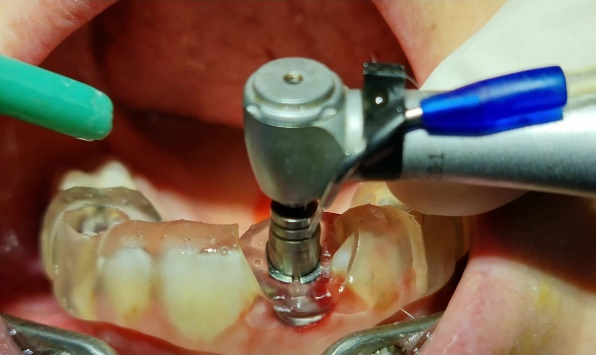
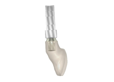
In order to ensure a precise and successful surgical outcome, the dentist used exocad to make a meticulously plan and designed the surgery guide. Once the design was complete, it was then expertly printed by state-of-the-art technology using AccuFab–D1s printer. The accurate surgery guide allowed for an unprecedented level of precision and control during surgery, ensuring that each step of the process was executed flawlessly.
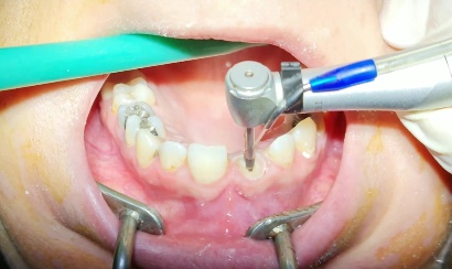
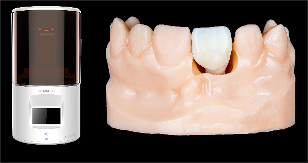
Make a Temporary Crown
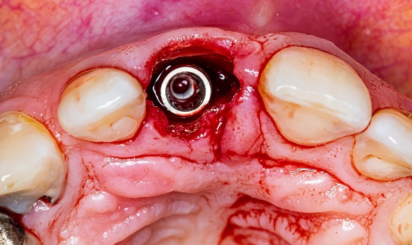
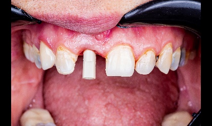
Right after surgery, they quickly designed a temporary crown and were able to use AccuFab-D1s printer with resin TN11 to create an accurate physical replica of the restoration designed in exocad – the entire process from beginning of the surgery to temporary teeth were all within just two hours. This allowed them to quickly and easily fit the new restoration into place with minimal discomfort for their patient.
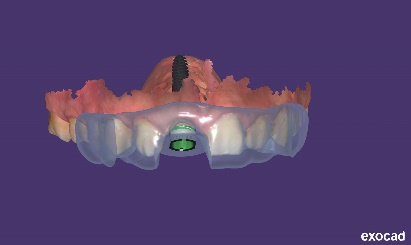
Scan and Design
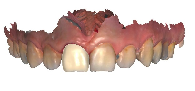
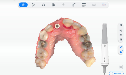
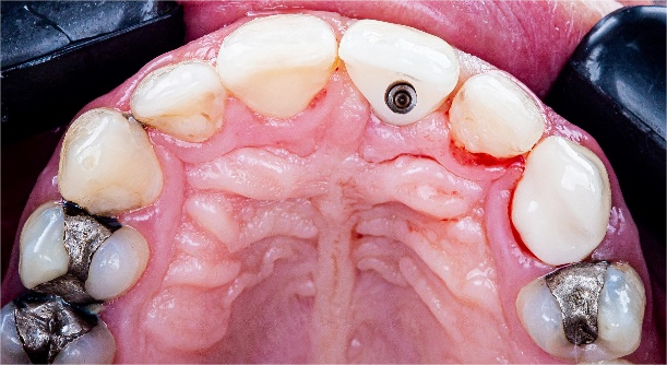
Two months later, the patient returned with good gingival health and successful osseointegration, the second stage of the treatment begin with an intraoral scan to capture the intraoral data.
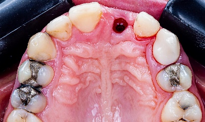
By using intraoral scanning technology, Drs. Khalaf and Hany were able to capture detailed information of the patient’s teeth without needing traditional impressions or molds. The intraoral scan data captured by Aoralscan 3 is accurate and life-like. These data were then used for further design.
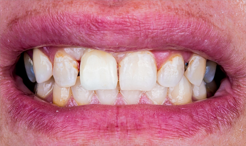
Place Final Restoration in Patient’s Mouth
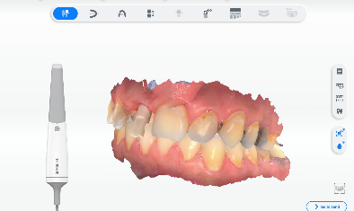
A model was 3D printed using AccuFab-D1s for quality inspection, and all the misfits were adjusted at this time, including color, adjacency, occlusion, etc. Once everything was ready, the dentist delivered the final zirconia restoration to the patient who expressed great satisfaction with the outcome.
Comments from Dentists
This case study highlights just how powerful digital technology can be when it comes to improving outcomes for patients while also making daily work easier for busy dental professionals like us. As these technologies continue to evolve and become even more advanced, we can expect even greater benefits for both patients and practitioners alike! Go digital with Shining 3D.