 ENG
ENG


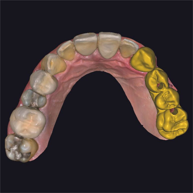
Table of Contents
The utilization of digital dental technology can significantly enhance the comprehensiveness of diagnoses, accuracy of final restorations, and overall efficiency of dental workflows. In today’s case study, we focus on Luis Estuardo Pacheco, an experienced dentist from Guatemala, who has established his own dental studio with the mission of assisting patients in restoring their smiles and completing the partial oral rehabilitation.
Case Profile
The patient, age 45, presented with multiple missing teeth and several deteriorated old restorations. Seeking an affordable solution, the patient expressed a desire to restore their teeth.
Data Collection
Initially, the dentist collected all necessary information during the patient’s first visit, including a panoramic radiograph, skull data, intraoral scan data, and face scan data. Armed with this comprehensive dataset, the dentist was able to analyze the case thoroughly, consider various treatment options, and select the most suitable one for the patient.
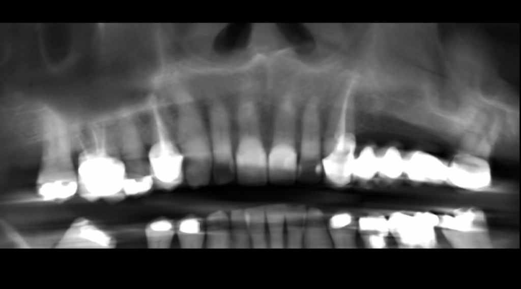
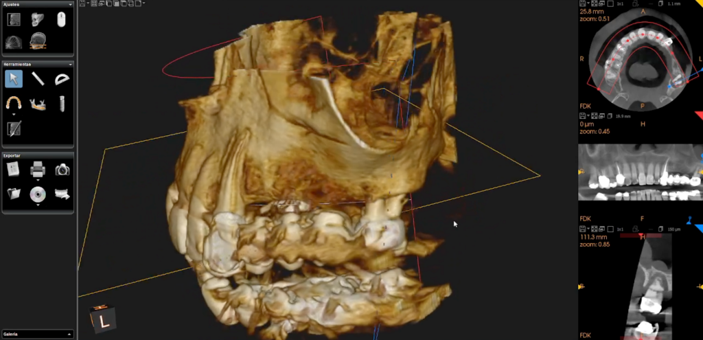
Fig 1,2: Panoramic radiograph & Analysis skull data and bone mass
Treatment Plan
Considering the patient’s budget constraints, the dentist decided to proceed with the restoration of the upper jaw for the first stage of treatment. The treatment plan involved the removal of the old pontic restoration in the #2 area and the placement of a single crown on tooth #23. Implant restorations were used for teeth #24-#26. It was noted that tooth #46 was missing, and the patient was aware that it would require future attention.
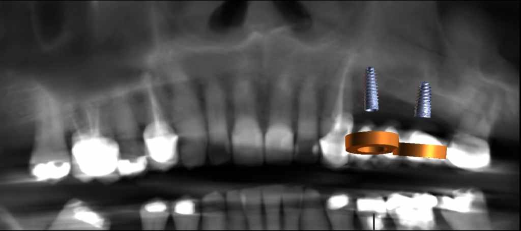
Fig 3: Treatment plan: two implant bodies were planned.
Treatment Process – Design and Print Surgical Guide
Utilizing the intraoral scan data acquired by the SHINING 3D Aoralscan 3, the dentist designed a surgical guide using dental software. This guided the surgical procedure by determining the position, depth, and angle of the two implant bodies. The designed surgical guide was then sliced using Accuware and printed with AccuFab-L4K using resin SG01.
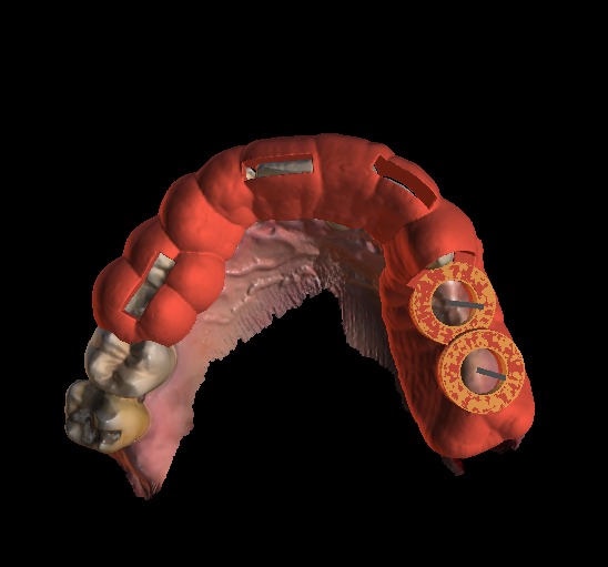
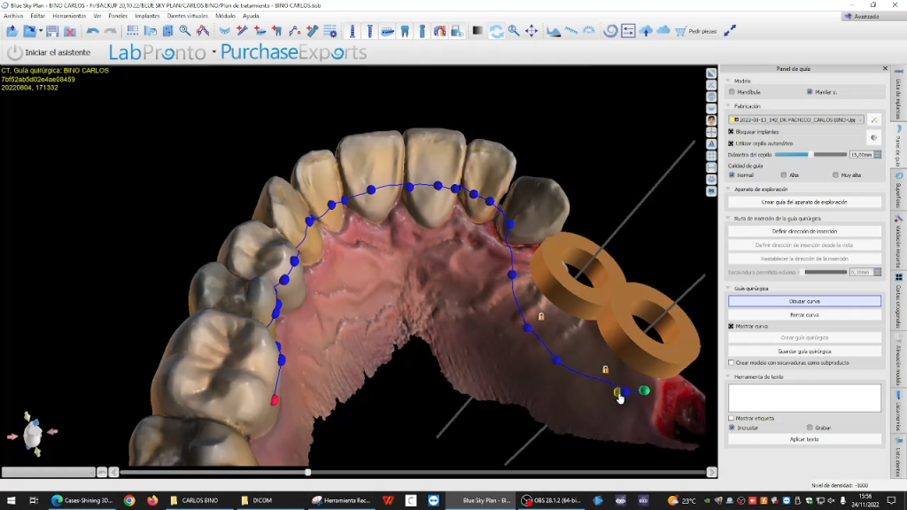
Fig 4,5: The design process of surgical guide.
Treatment Process – Implant Surgery
During the surgery, the surgical guide proved instrumental in ensuring the precise placement of the implant bodies. Following implant surgery, two healing caps were utilized to shape the emergence profile.
Treatment Process – Intraoral Scan
Two months later, the patient returned for upper restorations. The gingiva displayed healthy plumpness and a favorable emergence profile. The dentist captured intraoral data using Aoralscan 3. Initially, a scan without the scan body was performed to obtain clear emergence profile data. Subsequently, the scan body was installed, and a second scan was conducted to capture the scan body data for alignment in exocad. Occlusion data was obtained thereafter.
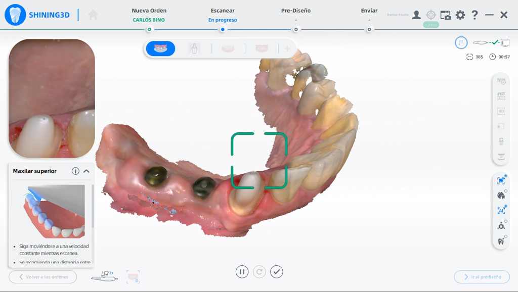
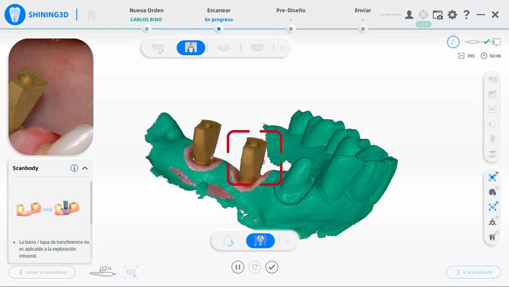
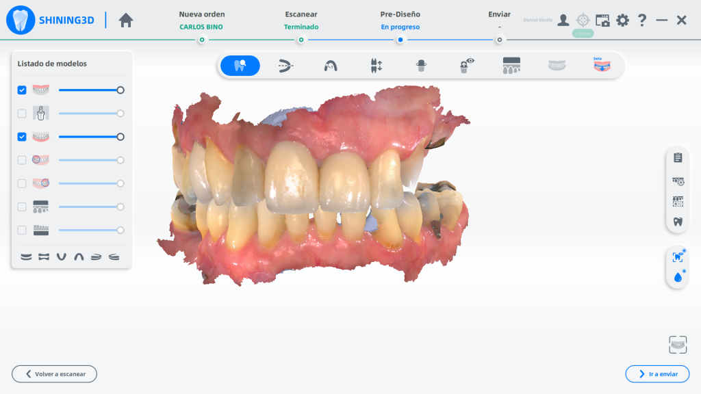
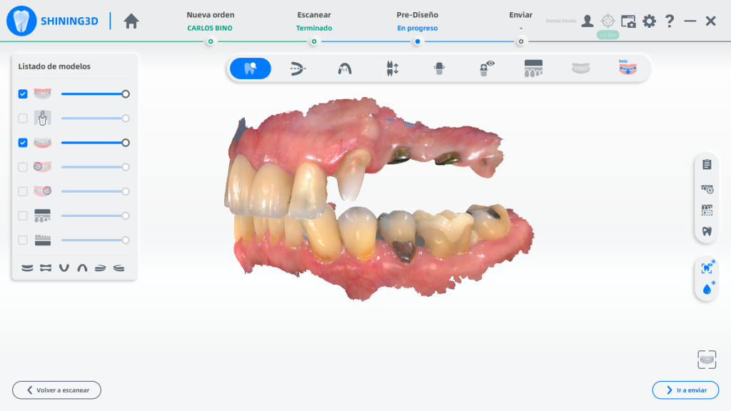
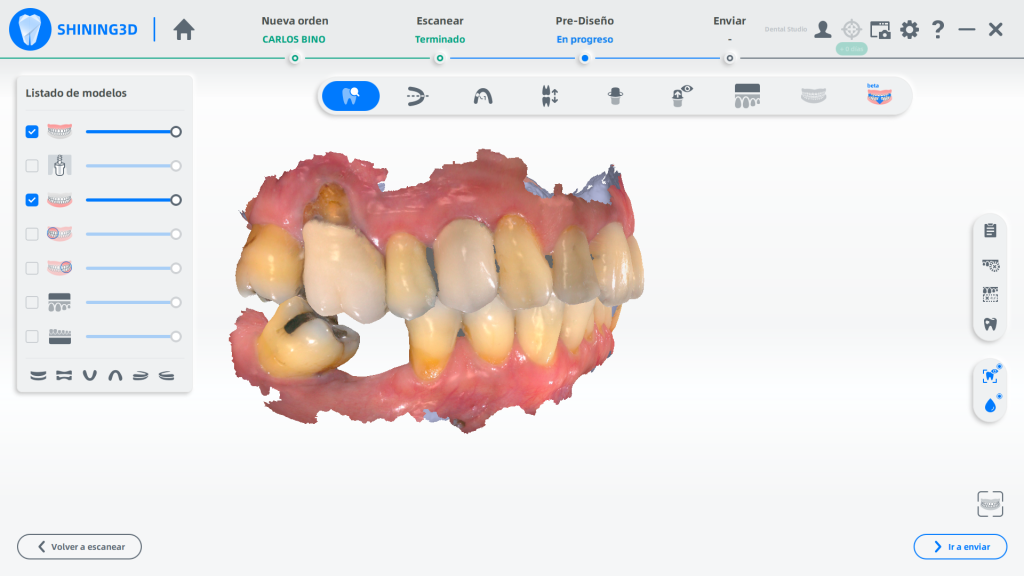
Fig 6,7,8,9,10: The intraoral scan process using Aoralscan 3.
Treatment Process – Restoration Design in exocad
The collected data, along with the face scan data, was imported into exocad, where the design process involved creating four teeth—a crown and an implant bridge.
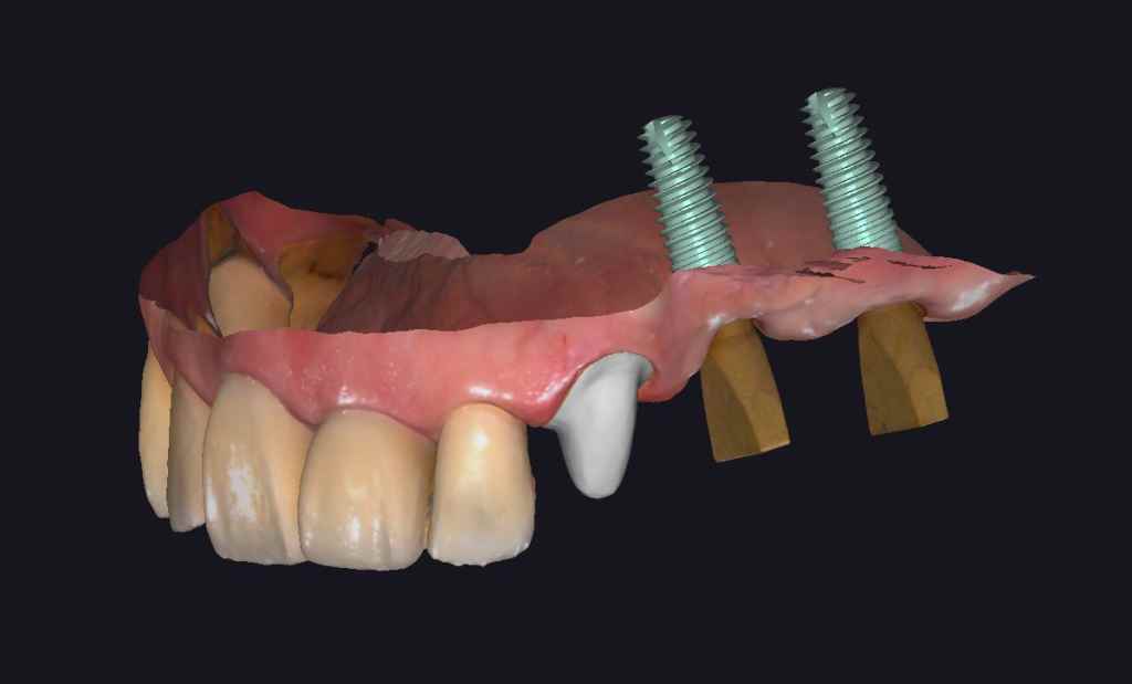
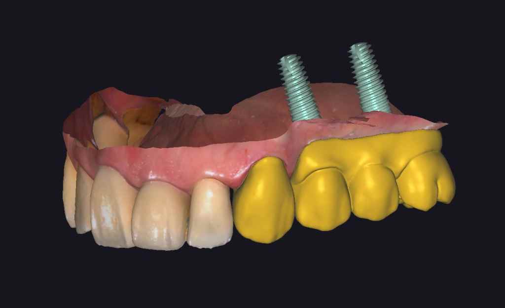
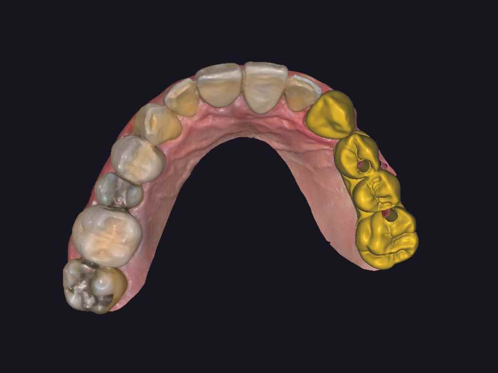
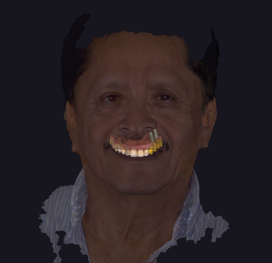
Fig 11,12,13,14: The design process in exocad.
Treatment Process – Model Print
The restorations were made of zirconia. A printed model was utilized for quality inspection, ensuring a precise fit of the prosthesis. Any issues identified during the inspection, such as adjacency, occlusion, or morphology, were adjusted at this stage.
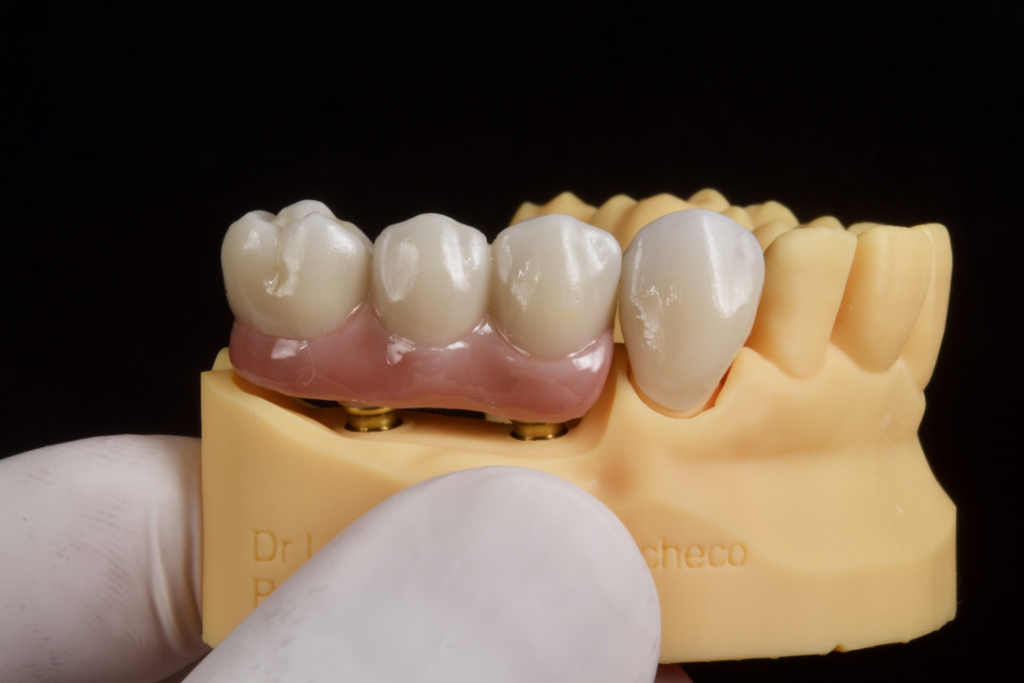
Fig 15,16: The final restorations were inspected in printed model.
Treatment Process – Teeth Delivery
During the final appointment, the dentist affixed the restorations securely in the patient’s mouth. A seamless fit was achieved between the abutment and the restorations, with optimal occlusion, adjacency, and lifelike color. The patient expressed high satisfaction with the final outcome.
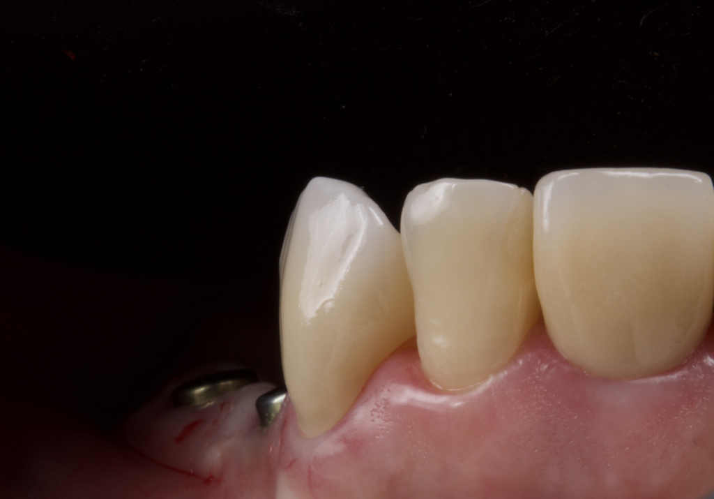
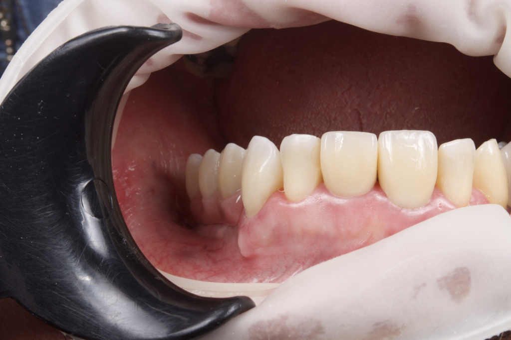
Fig 17,18: The restorations were placed with almost no adjustment.
About Author
- Dr. Luis Estuardo Pacheco
- DDS., MSc.
- CEO at Dental Studio, Guatemala.
- Senior CAD/CAM consultant, Scientific Team Lab(STL), 3D Labs CAD/CAM Laboratories, Guatemala.
- Senior consultant in oral and maxillofacial radiology at 3D Xray, Guatemala.
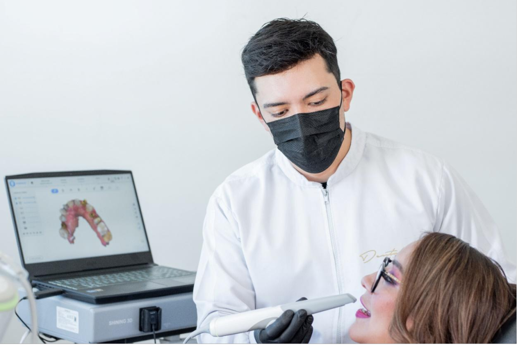
Fig 30: Dr. Luis Estuardo Pacheco