 ENG
ENG


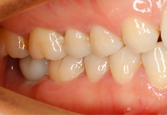
Table of Contents
Case Information
The 51-old female patient complained of the molar loss for half a year.
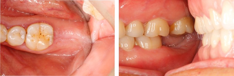
Diagnosis: No.47 tooth loss
Treatment: No.47 tooth implant, real-time temporary crown restoration and final restoration completed in three months.
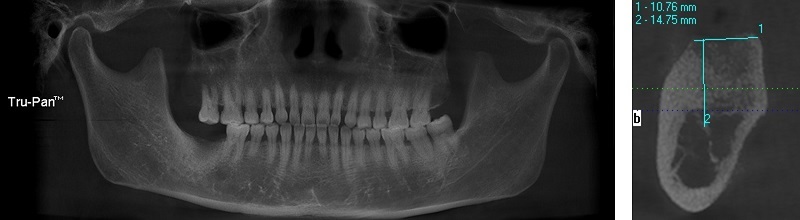
Preoperative Examination
A dental cone beam CT X-Ray scan was performed before surgery. The result showed that the target location has enough bone mass for the implant.
Implant Surgery
Prepared the cavity, and embedded the Nobel CC 5.0x10mm implant.
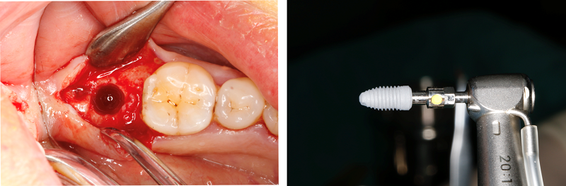
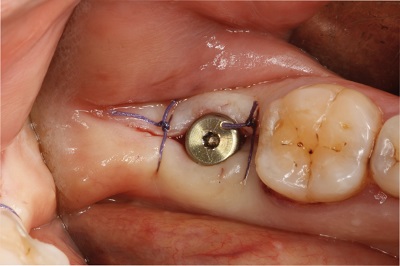
Implant Surgery Completion
The implant was embedded in the correct location and was in a stable status at the initial period.
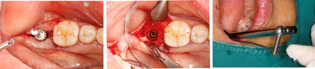
Scan Body Locating
Insert the sterilized scan body in the patient’s mouth, and make sure that it was correctly and completely located. It is possible to do an X-ray examination when necessary.
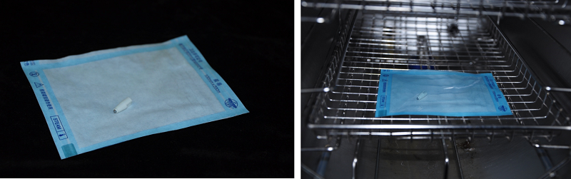
How To Sterilize Scan Bodies?
- Soak the scan body in liquid disinfectant for 5 minutes.
- Put the scan body in a sterilized vacuum bag.
- Sterilize the bag in the disinfection cabinet under 121℃ and 1.5 atmospheric pressure for 15 minutes.
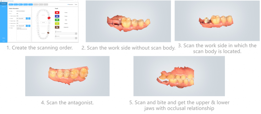
Intraoral Scanning
Use Shining 3D Aoralscan to scan the intraoral environment of the patient, which took about 7 minutes.
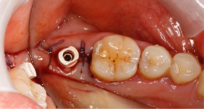
Healing Abutment Wearing
After scanning, put the healing abutment in the mouth while the temporary crown is being made.
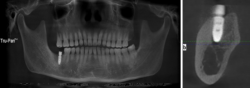
Postoperative CT Examination
After surgery, the dentist performed a dental cone beam CT X-Ray scan. The result showed that the implant was embedded correctly.
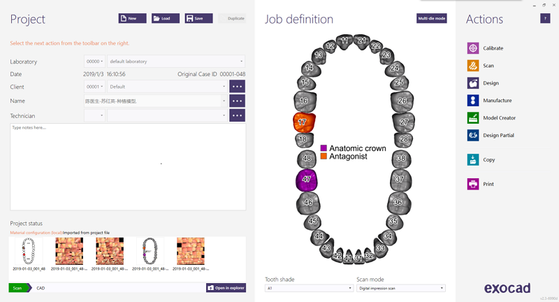
Intraoral Data Import into Exocad for Design
Import the intraoral data into Exocad and check the job definition.
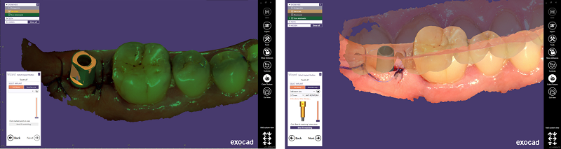
Scan Body Matching
Selecte the corresponding implant type in exocad and match the scan body position.
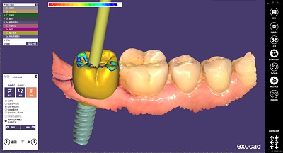
Temporary Crown Design
Design the temporary crown and the gingiva contact as required in exocad.
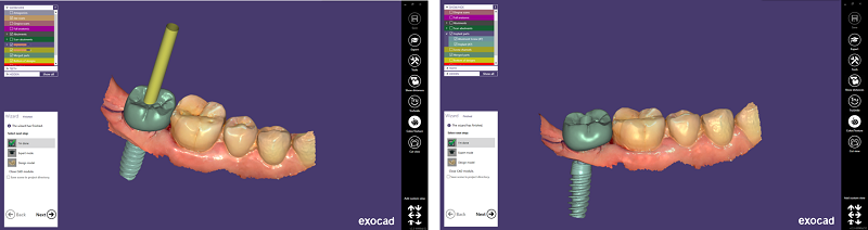
Design Completion
Complete the design of temporary crown in exocad.
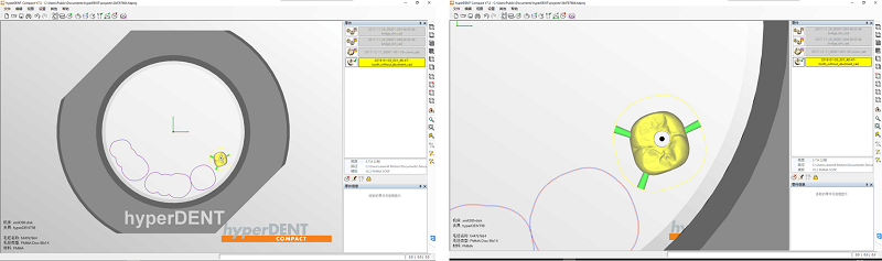
CAM Data Planning
Import the temporary crown CAD data into the CAM software and export the milling data after planning.
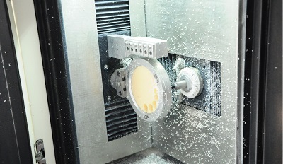
CNC Milling
It took about 20 minutes to mill the crown.
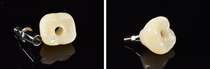
Milling Completion
Finish resin crown milling, stick the crown onto the base and implant temporary crown was formed.
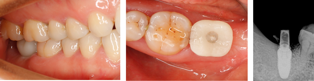
Try-in
Try the temporary crown in the patient’s mouth. The X-ray examination result indicated the correct crown location, suitable abutment, and slightly contacted occlusion. The clinic remodeled the gingiva during the temporary resin crown period.
Case Support

Dentist Introduction
Chen Qingsheng
Associate chief physician. Master’s degree in implant dentistry. Director of the oral implant department in Hangzhou Dental Hospital (Chengxi branch). Member of Chinese Stomatological Association Restoration Committee, Chinese Stomatological Association Aesthetic Restoration Committee. Standing director of Chinese Association of Aesthetic Dentistry. Prize winners of various national implant competition.
Comments from Dr. Chen
“Aoralscan is accurate, fast, efficient. With it the dentist can accurately acquire the intraoral information. Satisfactory restoration can be made with the use of equipped professional software for design and milling machine. This digital procedure reduces the chairside operation time and provides patients better experience. Hope it can be widely used.”
Benefits of a chairside temporary implant restoration with Aoralscan
Same-day digital dentistry
It only takes 1 to 1.5 hours to create a temporary crown using chairside digital dentistry. Hence, the patient can wear the temporary crown after implant surgery on the same day.
Better experience for the patient
Scanning the mouth only takes 5 to 7 minutes with the AoralScan and does not require any powder. Plus, it eliminates the discomfort that patients can feel in traditional, physical impression molding.
Good for the implant result
Wearing the temporary crown right away is good for gingiva regeneration, which leads to a better implant restoration result.
Easy operation for the dentist
It’s much easier for the dentist/assistant to get a digital impression with the Aoralscan than to get a physical impression in a traditional way.
Enhanced patient-dentist communication
Finally, realistic 3D intraoral data is a great tool for the patient to understand their situation and treatment plan.