 ENG
ENG


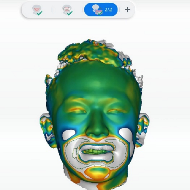
Table of Contents
Nowadays, traditional oral restoration is gradually being replaced by more convenient, rapid, and comfortable digital diagnosis and treatment methods. The integration of intraoral scanning and facial 3D scanning enables the reconstruction of digital virtual patient, providing personalized references for restoration design and predicting treatment outcomes. This enhances the overall treatment experience for patients.
Today’s case is presented by Dr. Wang Yuying (Fig1), who utilized various digital devices including the Aoralscan 3 and MetiSmile face scanner to successfully accomplish this aesthetic restoration case. The integration of the MetiSmile face scanner has significantly reduced the treatment time required for the entire procedure.
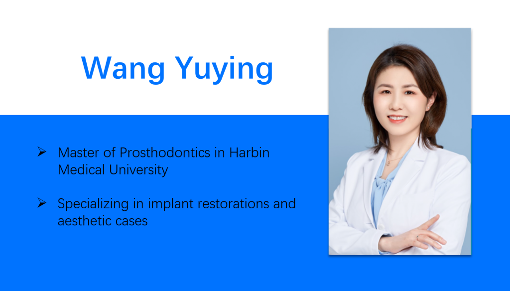
Fig 1:A brief introduction of Dr. Wang Yuying (Graduated from Harbin Medical University)
Case Profile
Chief Complaint:
The patient expressed concern regarding the retention of primary teeth in the anterior region of the maxillary arch, which has a significant impact on aesthetic appearance. Consequently, he sought consultation for implant-based restorative treatment (Fig 2).
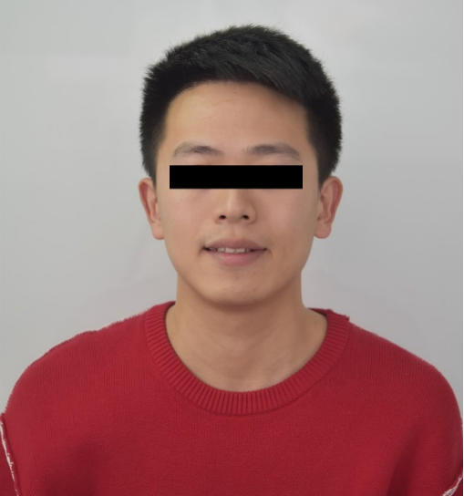
Fig 2: The front face photo before treatment.
Medical History
The patient presented with retained primary teeth bilaterally in the anterior region of the maxillary arch, resulting in occlusal imbalance. This condition had led to frequent lip biting, impacting both aesthetics and comfort. The patient denied any history of systemic illnesses.
Intraoral Analysis
- There were cavities in the primary teeth on both sides, and they were too small, severely worn, and sharp.
- The permanent central incisors in the maxillary arch displayed incomplete enamel development and discoloration.
- The lateral incisors were abnormally undersized.
- There were problems with teeth misalignment, malocclusion, dental rotations, and big interdental spacing.
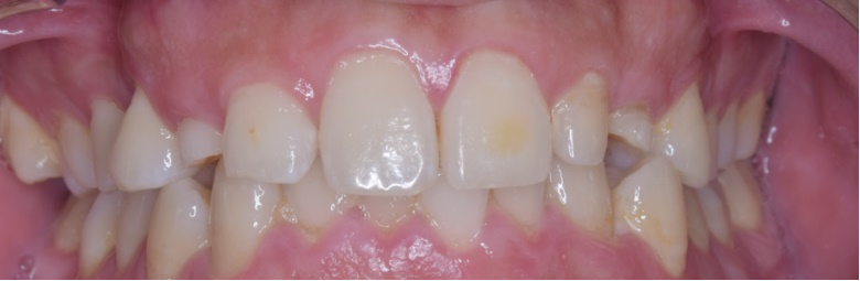
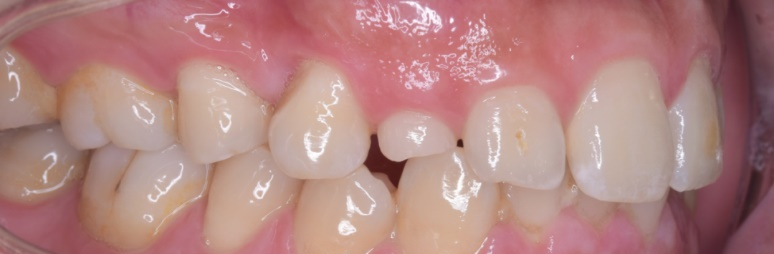
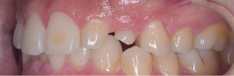
Fig 3-5: The intraoral photo before treatment
CBCT Analysis
Upper left 3 tooth: primary tooth’s root was absorbed, the root length was less than 1 mm.
Upper right 3 tooth: primary tooth’s root was 14 mm long with sufficient bone mass.
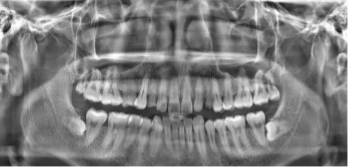
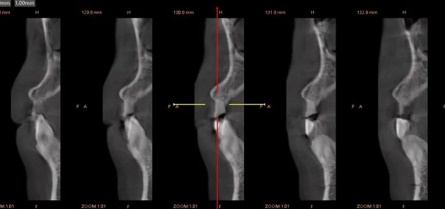
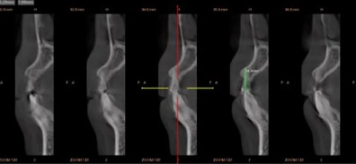
Fig 6-8: The CBCT data before treatment
Treatment Plan
Data Capture
Facial Photos Before Treatment
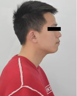
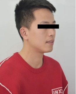
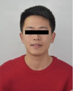
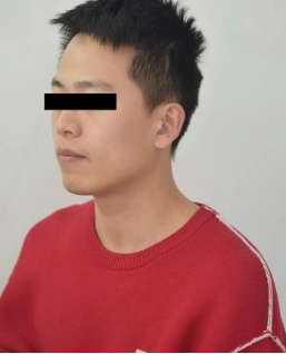
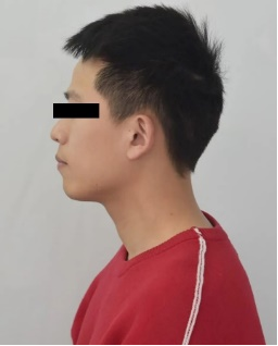
Fig 9-13: Dentist took several photos to record the 2D facial information from different angles.
Intraoral Scan Data Captured by Aoralscan 3 Before Treatment
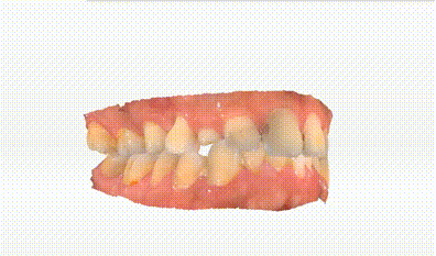
Fig 14: The intraoral data captured by Aoralscan 3
Facial Data Captured by MetiSmile Before Treatment
The MetiSmile 3D face scanner, manufactured by SHINING 3D, was utilized to acquire pre-operative facial information. This advanced technology enables automatic and precise alignment of facial data with intraoral scan data, as well as extraction of lip lines. Consequently, it provides dependable data for accurate diagnosis, treatment planning, and analysis.
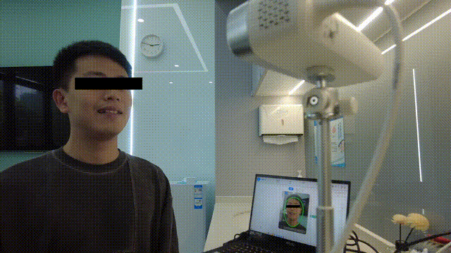
Fig 15:Facial data captured by MetiSmile
The Auto Alignment Between Extracted Facial Data and Smile Facial Data
The alignment result between extracted facial data and smile facial data can be visualized using a ribbon map.
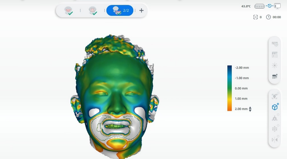
Fig 16:The alignment between extracted facial data and smile facial data
The Auto Alignment Between Extracted Facial Data and Intraoral Scan Data
The alignment between facial data and intraoral data can be accurately achieved within the face scanner software, eliminating the need for third-party software.
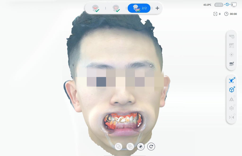
Fig 17: The auto alignment between extracted facial data and intraoral data
Lip Line Extraction
The extraction of the lip line can be automatically performed during the pre-design stage, and it can also be adjusted manually.
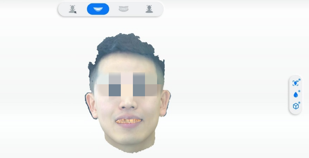
Fig 18: The lip line extraction in the pre-design stage
Design and Treatment
Aesthetic Design
Based on the collected data, the dentist proceeded with an aesthetic design, incorporating dynamic occlusion as a reference. The mock-up models were swiftly printed using AccuFab-D1s at chairside within 15 minutes (Fig 19). Subsequently, teeth preparation guides and resin silicone rubber plates were made based on the printed mock-up model (Fig 20).
Video 1:The aesthetic design process of mock-up
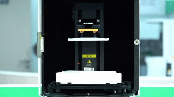
Fig 19: The printing process of mock-up model.
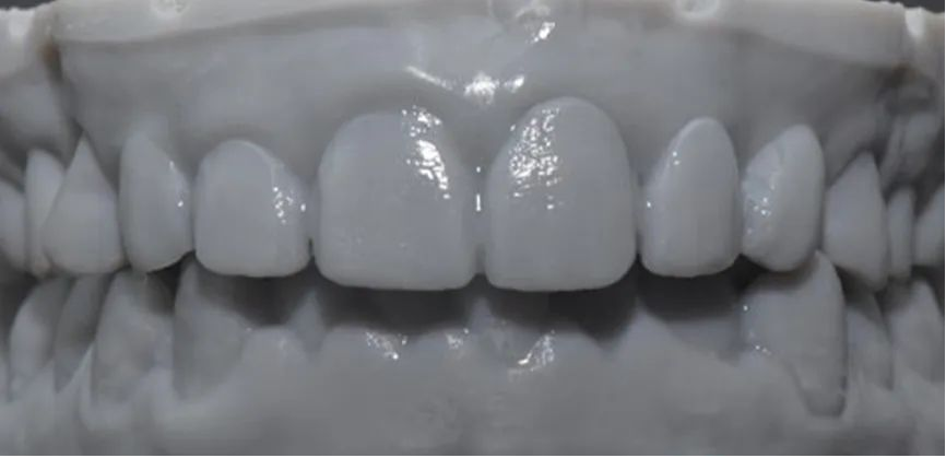
Fig 20:The intraoral photo of mock-up.
Teeth Preparation
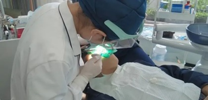

Fig 21, 22: The dentist performed teeth preparation under the guidance of a teeth preparation guide, ensuring accuracy throughout the process.
Video 2: The intraoral scan data after teeth preparation.
The intraoral scan data was imported into exocad, and the final restorations were designed based on the mock-up morphology. Throughout the design process, integration of intraoral scan data, face scan data, and CBCT data ensured utmost accuracy and suitability of the final restorations for the patient.
Video 3: Check the aesthetic design of the final restorations in exocad
Fixing Restorations
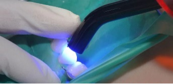
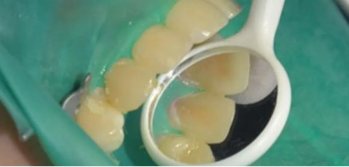
Fig 23,24: Fixing the #13 all porcelain crown.
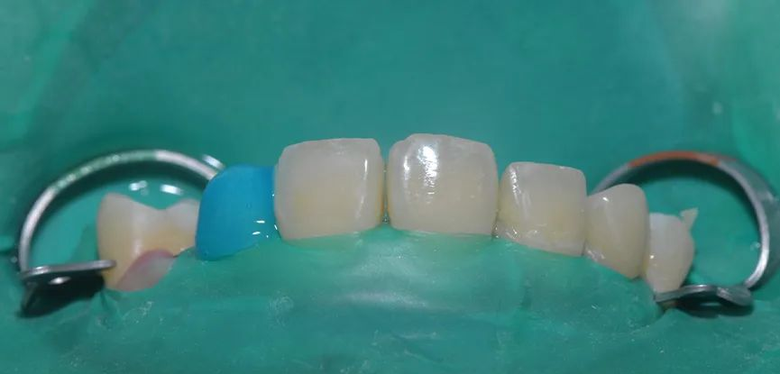
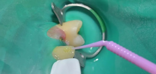
Fig 25,26: Fixing the #22 ultra-thin veneers.
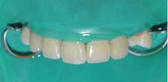
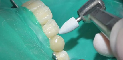
Fig 27, 28, Forming composite resin at mesio-incisal angle of #11,#12.
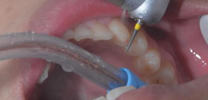

Fig 29,30: The occlusion adjustment.
Final Effect
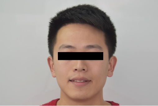
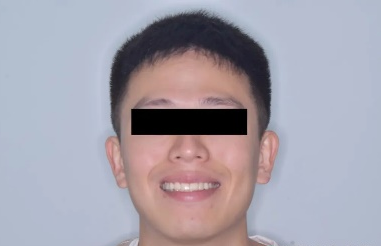
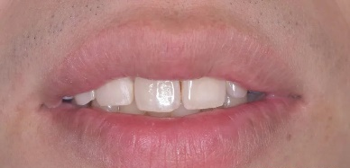
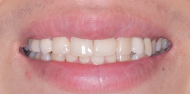
Fig 31-34: Before treatment (Left) & After treatment (Right)
Summary
In this case, the patient had been suffering from maxillary anterior tooth defects for an extended period of time, which significantly impacted his overall oral health. Considering the treatment effectiveness and the limitations on the patient’s visits, we utilized MetiSmile face scanner to construct a precise 3D face model and Aoralscan 3 intraoral scanner to create an accurate teeth digital model. With a virtual patient as a reference during the design process, a mock-up was processed for a comprehensive communication and planning confirmation before the teeth preparation begun. Visual diagnosis and treatment make patients more intuitive to understand the whole procedure and strengthens the communication between the dentist and the patient. As a result, the patient expressed satisfaction with the entire treatment process.