 ENG
ENG


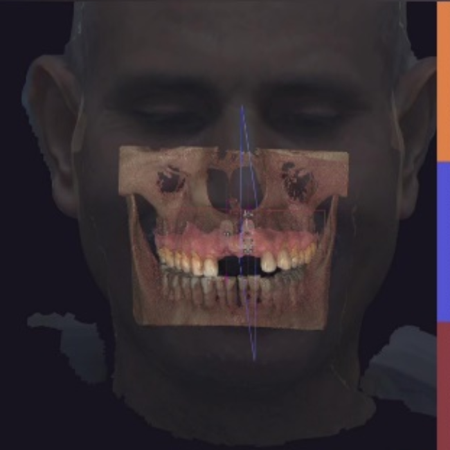
Table of Contents
The utilization of facial scanners has gained traction in the digital dental workflow in recent years. This technology employs advanced scanning techniques to digitally capture and construct a detailed 3D facial model. In a recent case study provided by Dr. Abdelrahman Khalaf and Dr. Kirollos Hany from the Faculty of Dentistry at Assiut University in Egypt, they demonstrated how the 3D facial scan data can be used to restore the anterior aesthetic zone for implant patients.
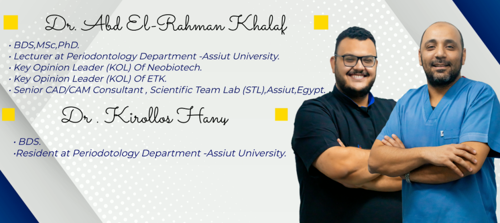
Fig 1: Dr. Abdelrahman Khalaf and Dr. Kirollos Hany
Case Profile
The male patient, who had lost his anterior teeth due to an accident, experienced issues with pronunciation, chewing, and outlook appearance, resulting in a decrease in his quality of life. Therefore, the patient visited Dr. Abdelrahman Khalaf and Dr. Kirollos Hany to have this issue fixed with long-lasting restorations.
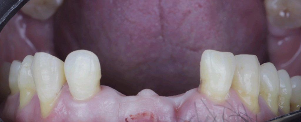
Fig 2: Before treatment: the patient lost two teeth, he was seeking restoration in the aesthetic zone.
Data Capture
Upon the patient’s arrival, Dr. Abdelrahman Khalaf and Dr. Kirollos Hany collected all the necessary data for a comprehensive diagnosis. First, they performed an intraoral scan using Aoralscan 3, an elegant scanner with high accuracy that can quickly capture, the upper, and lower jaws and occlusion data.
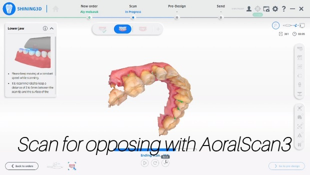
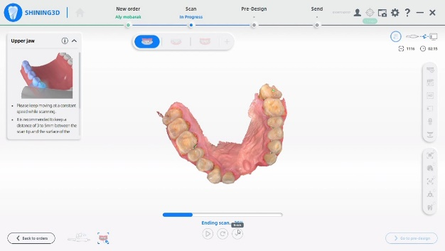
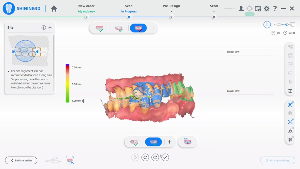
Fig 3-5: The intraoral scan data of the upper jaw, lower jaw, and occlusion.
Then the dentists captured the 3D facial data using MetiSmile, a 3D face scanner developed by SHINING 3D specifically for dental applications. The intraoral scan data and the face scan data can be automatically aligned in the software to create a virtual patient.
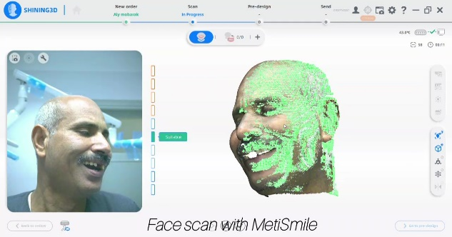
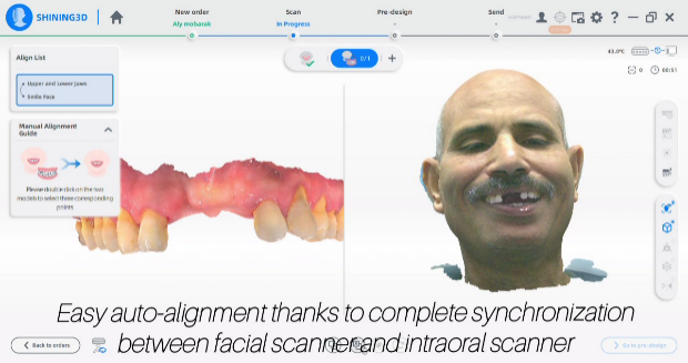
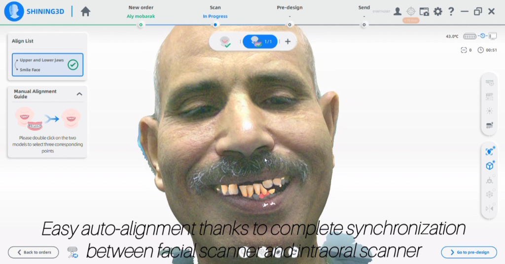
Fig 6-8: The dentist captured the patient’s facial data using the fixed scan mode, the alignment between intraoral scan data and face scan data is accurate.
Treatment Plan
With the help of comprehensive information, the dentist had a thorough discussion with the patient, considering the treatment outcome forecast, long-term usage, and the patient’s experience during treatment. The final treatment plan involves designing and printing restorations in advance. Then, an implant surgery is performed under surgical plate guidance. Afterwards, the pre-prepared interim restorations are fixed into the patient’s implant position to avoid waiting time.
During this process, three-dimensional visualization for diagnostics and treatment planning is crucial and necessary for prosthetic rehabilitation, especially in implant cases. The creation of a virtual patient, by combining data facial scanners, intraoral scanners, and CBCT, can streamline diagnostic and design procedures.
This treatment plan relies heavily on advanced digital technology for capturing all necessary information and on dentists’ precise surgical skills to accurately place the implant body as planned.
Design Process
Dentists designed the two anterior teeth, #11 and #21 in exocad. Considering that esthetics are highly subjective and influenced by various factors, employing a virtual facial model enables effective collaboration between the clinician and patient for personalized treatment planning.
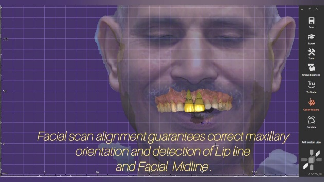
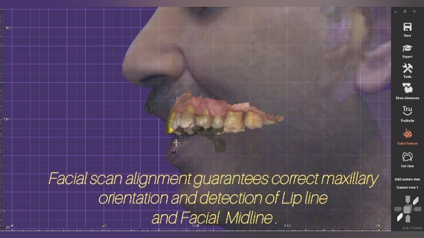
Fig 9, 10: the facial scan alignment guarantees correct maxillary orientation and detection of the lip line and facial midline.
Except for the interim restorations, the dentists also designed the implant surgical planning with the help of a facial model.
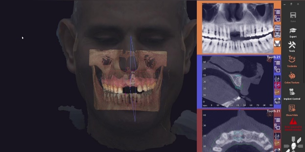
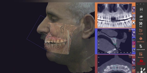
Fig 11, 12: The surgery planning for the implant was done in exocad.
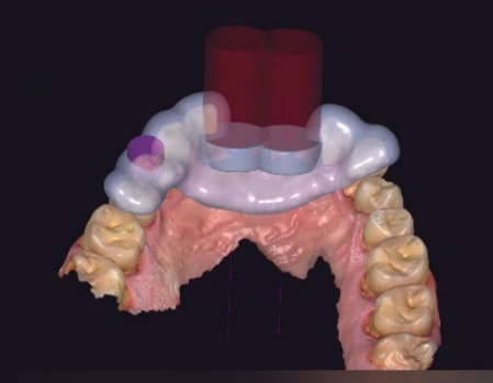
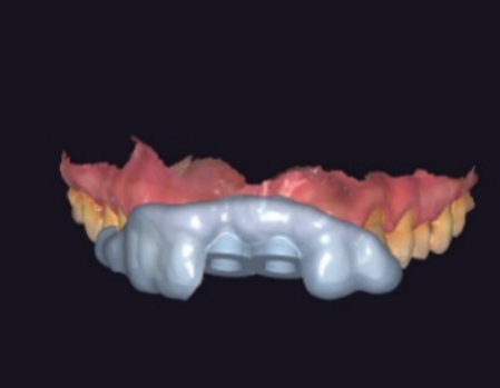
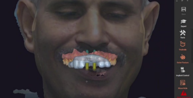
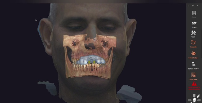
Fig 13,14: The surgical guide was designed in exocad.
Implant Surgery Process under the Surgical Guidance
Then the dentists printed the implant surgical guide using AccuFab-D1s, an accurate printer with super-fast speed. With the help of this surgical guide, dentists can accurately place the implant scan body in the right position with suitable depth and angle.
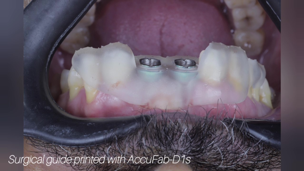
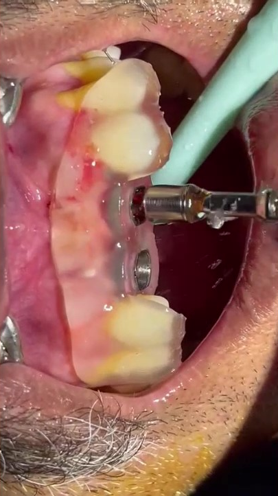
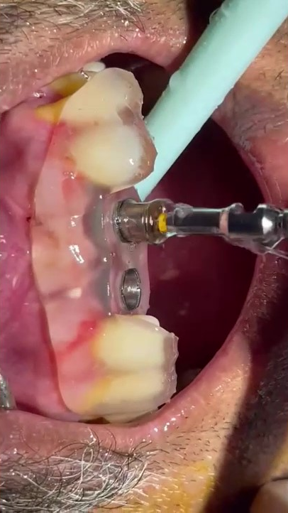
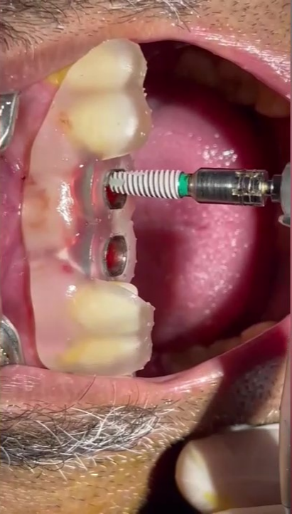
Fig 15-18: the implant surgical process.
Immediate Interim Restorations
After that, the interim resin restorations were placed in the patient’s mouth on the same day. They will be used while the patient waits for osseointegration and final restorations. allowing the patient to chew and pronounce normally.
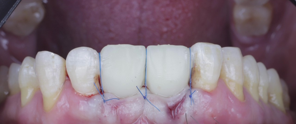
Fig 19: the interim restorations were placed in the patient’s mouth after implant surgery.
The Comments from Dr. Abdelrahman Khalaf and Dr. Kirollos Hany
MetiSmile is a rapid and non-invasive tool that can be utilized in multiple facets of dental care. In implant cases, it can serve an invaluable role by creating a virtual patient, improving the accuracy of diagnosis and surgery planning, and helping us achieve the best treatment results for patients.