 ENG
ENG


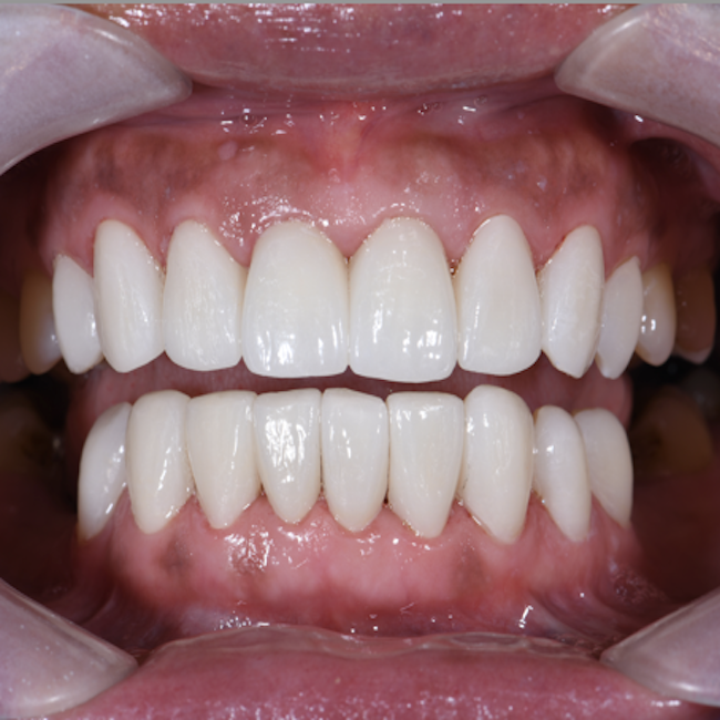
Table of Contents
With the development of digital technology, traditional anterior aesthetic restorations have been replaced with digital impressions, digital mock-ups, and temporary teeth. During the process of restoring anterior teeth, which requires high aesthetics, the shape of the temporary teeth can be replicated almost 100% using digital technology.
Today’s case comes from Dr. Gu Kaikai (Fig. 1) and technician Wang Zhilei (Fig. 2), who have been working together for years to provide beautiful restorations and a better smile to patients. In this case, Dr. Gu and technician Wang performed aesthetic anterior veneer restorations using the Aoralscan 3 intraoral scanner, an AccuFab-D1s printer, and a 3D facial scanner to achieve a presentable treatment.

Fig. 1: Gu Kaikai: Doctor of stomatology from Tongji University, specializing in minimizing aesthetic restorations; Deputy Medical Director of Hangzhou Dental Hospital.

Fig. 2: Wang Zhilei: Senior technician and trainer; Founder of Hangzhou Henyi Medical cooperation
Case Profile
The patient complained that the shade and shape of her anterior teeth were not beautiful enough (Fig. 3).
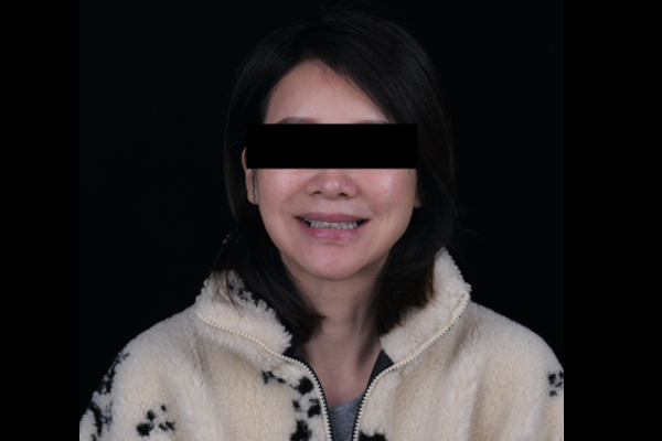
Fig. 3: The front facial view of the patient before treatment.
After clinical and imaging examination, the diagnoses were that there was a triangular space in the anterior tooth area, between #11 and #21. The teeth were in good health with no loosening, and no obvious malfunction of the gingiva (Fig. 4, Fig. 5, Fig. 6, Fig. 7, Fig. 8).
Based on the comprehensive communication with the patient, the ultimate treatment plan was customized with minimally invasive veneers, by which the patient could fix her issue and, at the same time, maintain as much of her own tooth structure as possible.
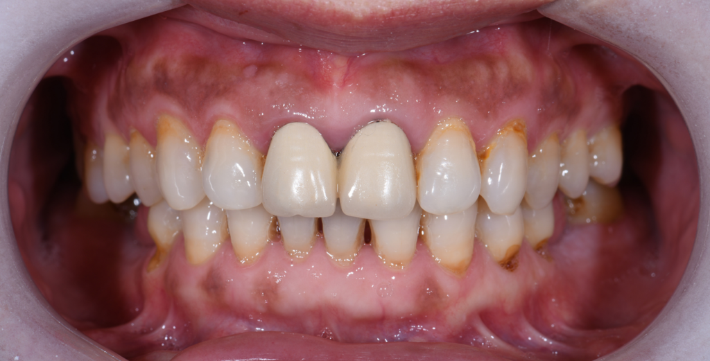
Fig. 4: The intraoral situation before treatment.
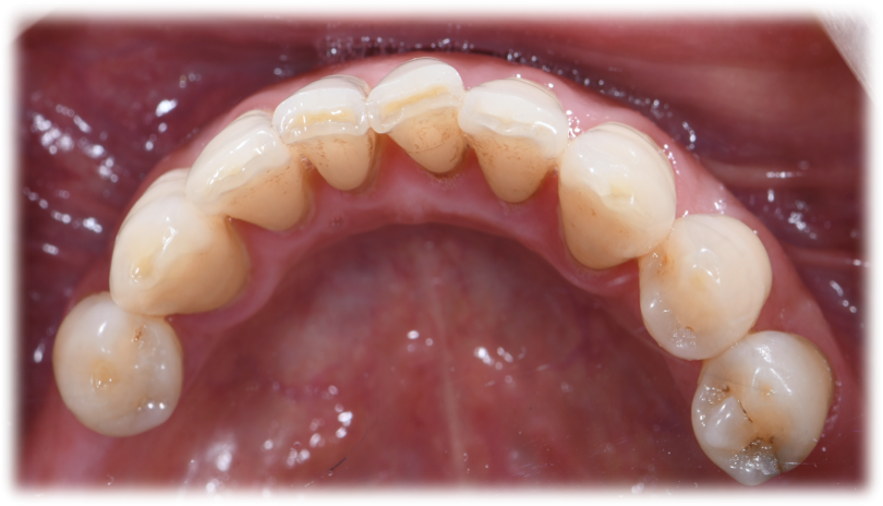
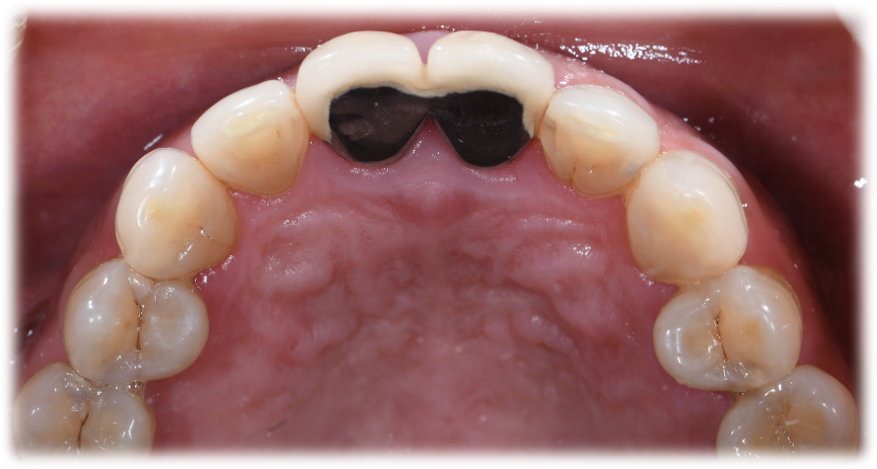
Fig. 5,6: The occlusal view of the lower jaw and upper jaw.
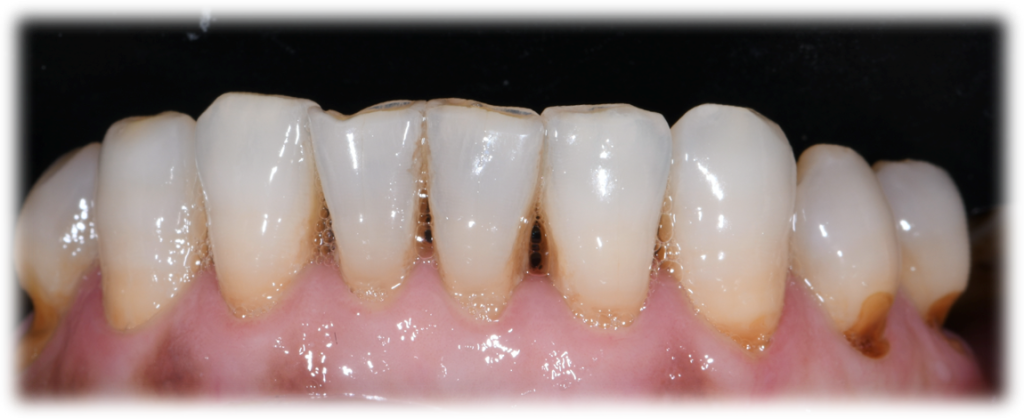
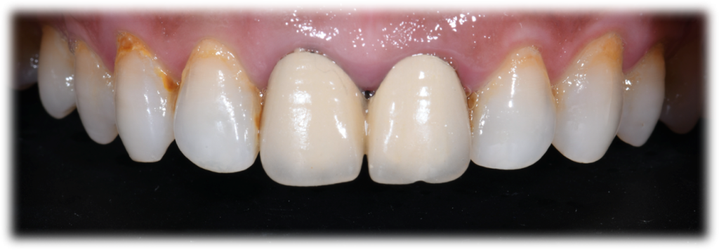
Fig. 7,8:the labial view of the upper jaw and lower jaw.
Data Collection Before Treatment
Comprehensive collecting of data before treatment aids in a smoother, easier, and more predictable plan. The intraoral data can be combined with CT and face scan data, which is significant to get a comprehensive and aesthetic design. This information also provides better communication between the dentist, patient, and technician, which in turn helps to get a better treatment outcome.
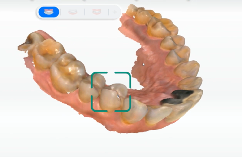
Fig. 9:Intraoral scan by Aoralscan 3: Upper jaw
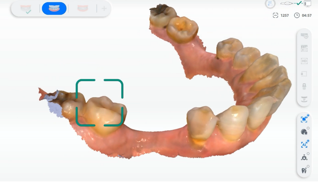
Fig. 10: Intraoral scan by Aoralscan 3: Lower jaw
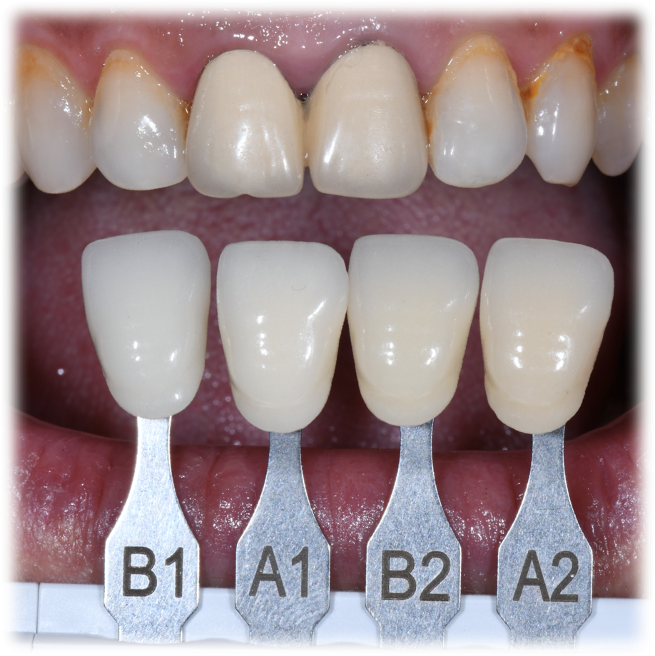
Fig. 11: The comparison between shade guide and the current tooth shade.
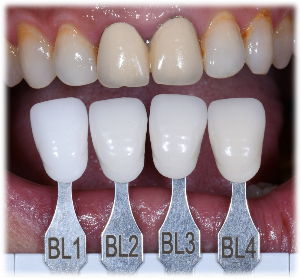
Fig. 12: The comparison between targeted shade and the current tooth shade.
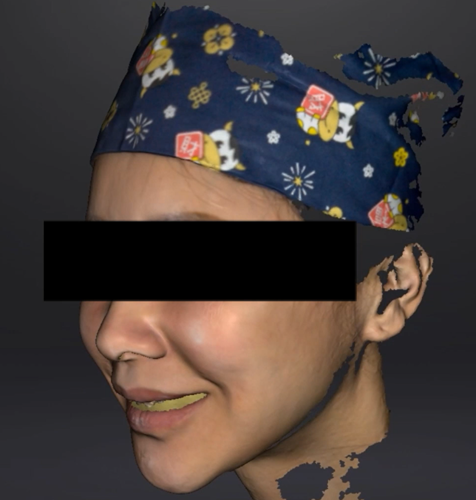
Fig. 13: The facial information collected by a face scanner.
During Treatment – Teeth Preparation
After collecting all the data, the dentist performed teeth preparation (Fig. 14, Fig. 15, Fig. 16, Fig. 17) and abutment shade match (Fig. 18, Fig. 19), and scanned again to obtain the information after teeth preparation (Fig. 20, Fig. 21).

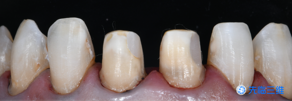
Fig. 14,15: The occlusal and labial view of the upper jaw after teeth preparation
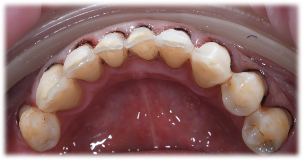
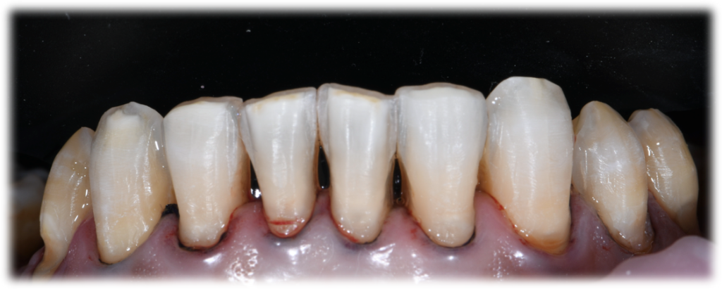
Fig. 16,17: The occlusal and labial view of the lower jaw after teeth preparation
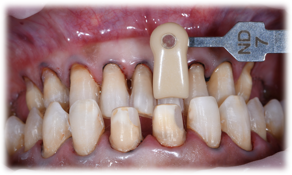
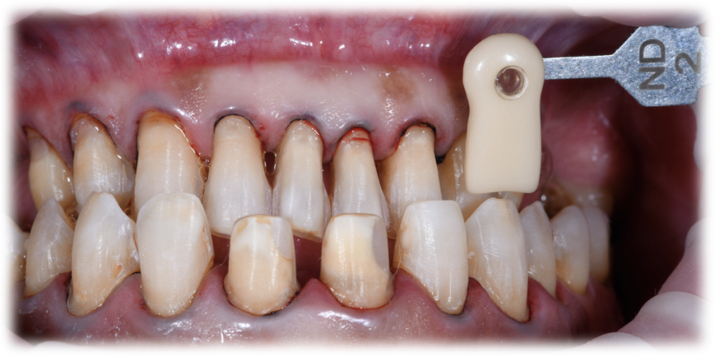
Fig. 18,19: The shade match of the abutment
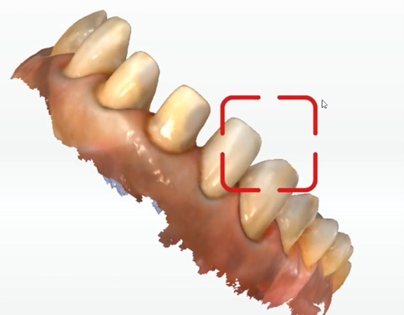
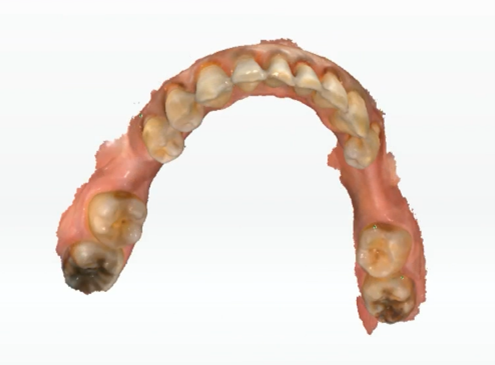
Fig. 20,21: The scan process after teeth preparation.
During Treatment – Computer Aided Design
After the teeth preparation, the mock-up was confirmed and the morphological scanning was performed, which could be used as a reference to complete the morphological design of the final restorations.
When designing the restorations, import the mock-up data to achieve patient satisfaction with the final prosthesis (Fig. 22). During the design, importing the facial scan data at the same time, simulating the delivery of the teeth in the patient’s mouth, and give a comprehensive evaluation about whether the designed teeth match the face shape and whether the protrusion of the dental arch is appropriate (Fig. 23).
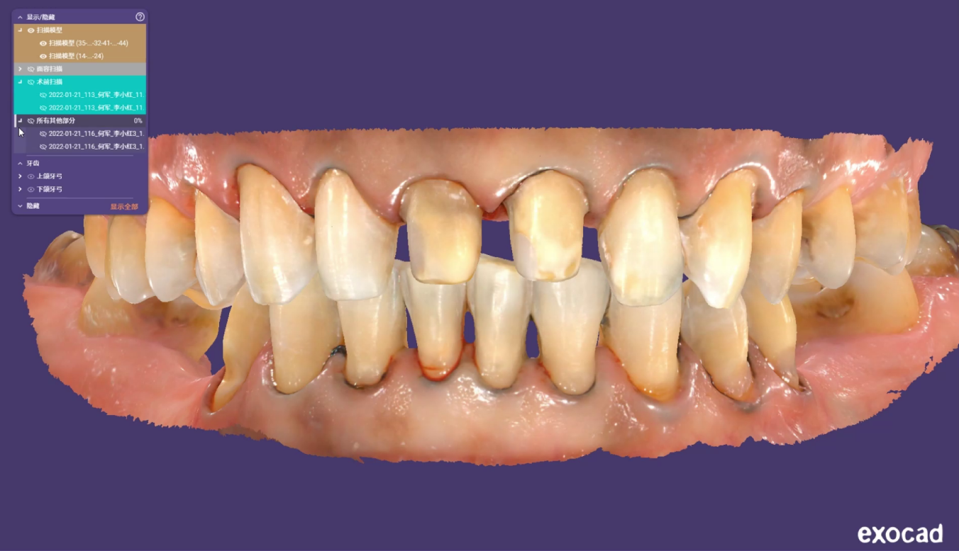
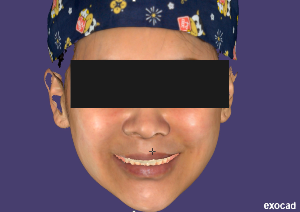
Fig. 22, 23: The design process.
During Treatment – Veneers Manufacturing and Model Printing
Manufacturing the veneers by the milling machine and printing the inspection model.
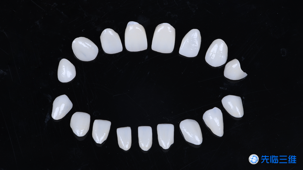
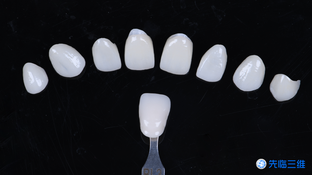
Fig. 24,25: The finished veneers.
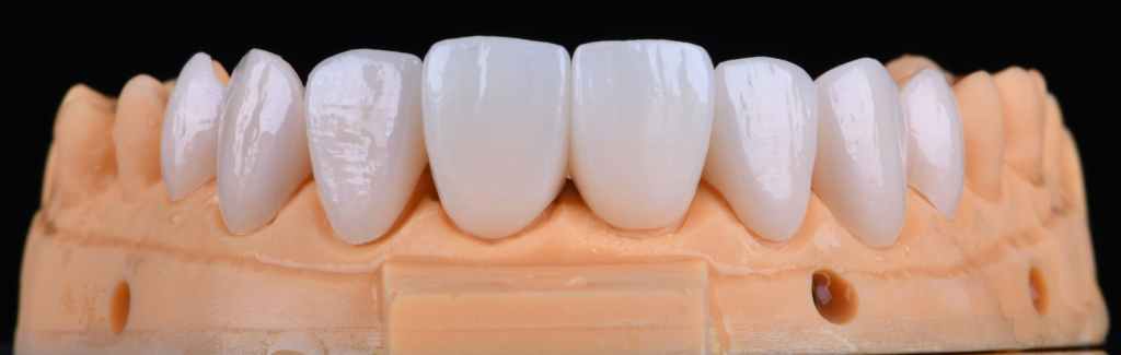
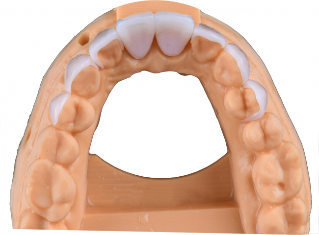
Fig. 26,27: The finished veneers of the upper jaw fitted well in the model.
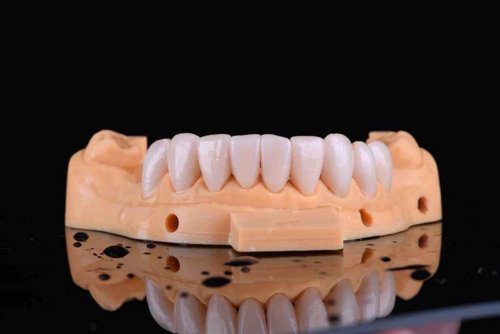
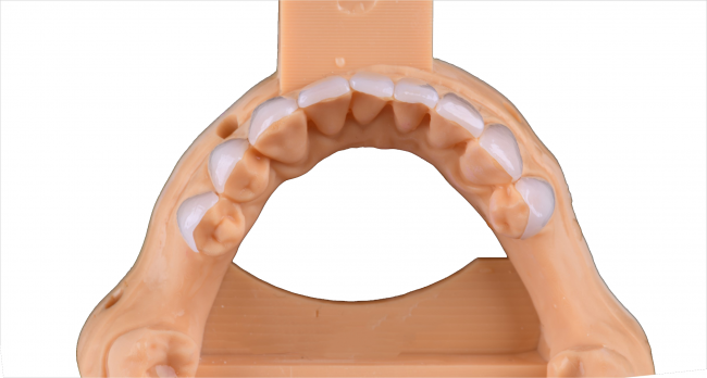
Fig. 28,29: The finished veneers of the lower jaw fit well in the model.
After Treatment
After the restorations were fixed permanently in the patient’s mouth, a harmonious and benign intraoral functionality was observed. And the final delivery of the aesthetic anterior veneers restorations is delivered.
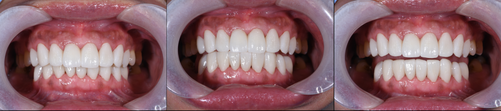
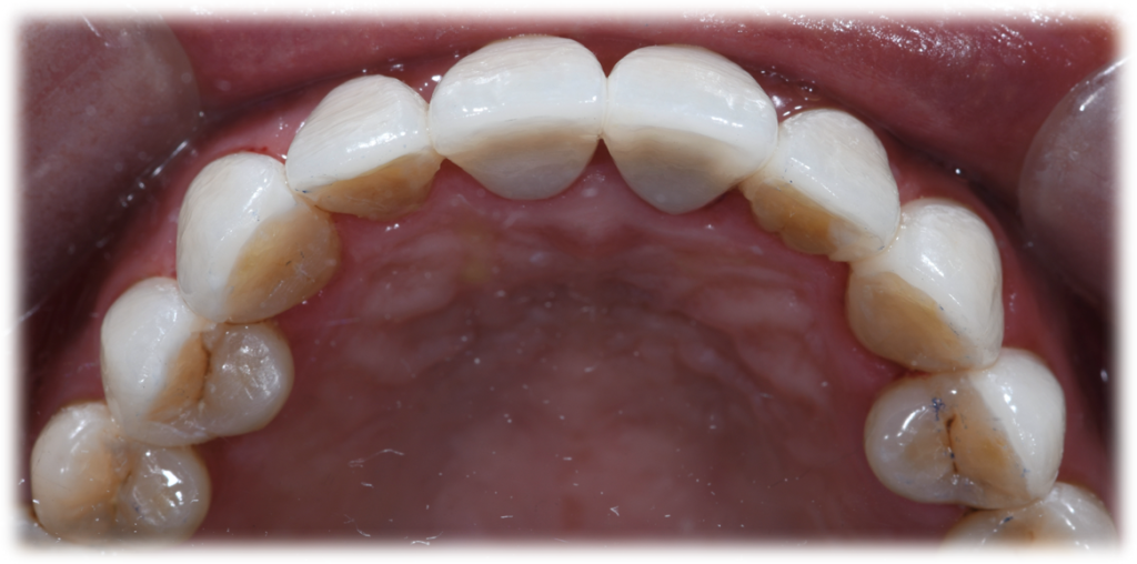
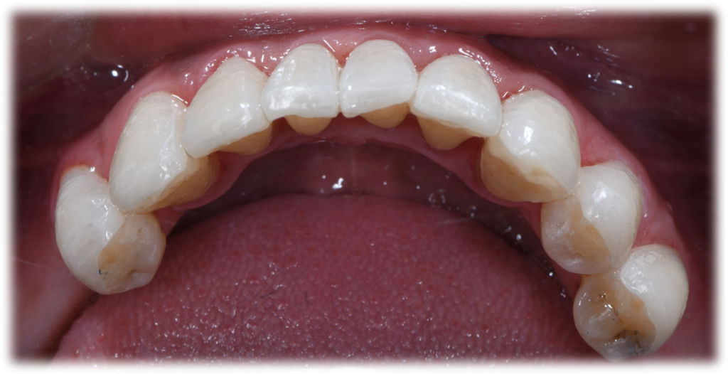
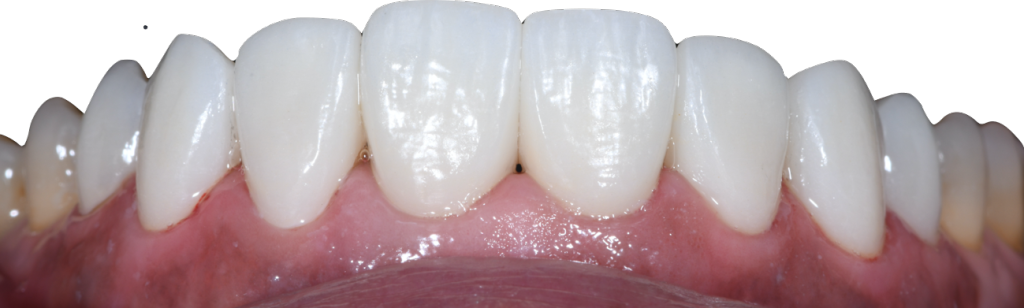
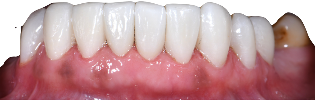
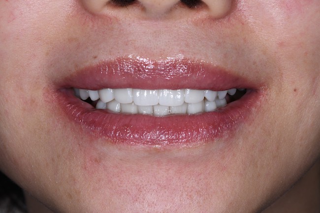
Fig 30-35: Final delivery of the aesthetic anterior veneers restorations
Summary
In this case, the patient was unsatisfied with the shape and shade of her previous teeth. Through aesthetic analysis and digital design, the dentist determined to combine the three-dimensional aesthetic design, the gingival height ratio, smile curve, and face shape ratio for a comprehensive production of digital aesthetic veneers. The patient was very pleased with the treatment experience and her final restorations.