 ENG
ENG


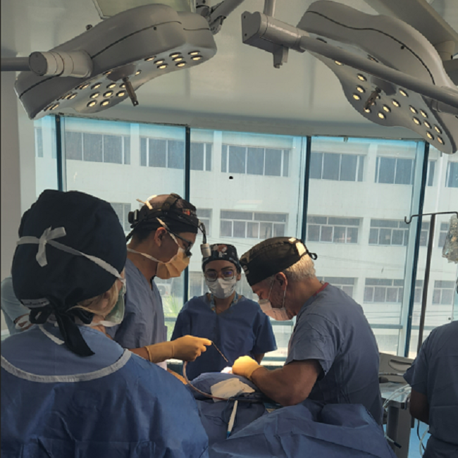
Table of Contents
Cleft lip and palate are severe congenital craniofacial malformations, with an incidence rate ranging from 1/2500 to 1/500 in newborns. The incidence rate is related to factors including geographical location, ethnicity, and socio-economic status. In Latin America, particularly in the Caribbean region, approximately one out of every 700 live births are affected by cleft lip and palate.
Today, we would like to present a case study of two neonates, Luna and Jesús, who were diagnosed with cleft lip and palate. They received comprehensive treatment under the guidance of Dr. Jadad, Dr. Sandra Cáceres M, Dr. Sergio Tinoco, Dr. Jorge Navarro and Harvard Medical Team.
Video 1: Rehabilitation of newborn babies with cleft lip and palate
The story originates from November 2022, when Dr. Jadad was invited to deliver a lecture at an academic conference held at the University of Sinaloa in Mazatlán, Mexico. During his presentation, he shared his clinical experiences using Aoralscan 3 and specifically highlighted its application in pediatrics through the use of the mini scan tip.
After listening to Dr. Jadad’s presentation, Dr. Sandra Cáceres M, an invited guest speaker, realized the tremendous potential of Aoralscan 3 and its mini scan tip in treating infants with cleft lip and palate. She suggested to Dr. Jadad the idea of using this device to scan the intraoral data of newborns as a foundation for subsequent treatment. As a result, Dr. Jadad and Dr. Sandra Cáceres M initiated their collaboration and started the treatment of the cleft lip and palate cases of the infants Luna and Jesús.
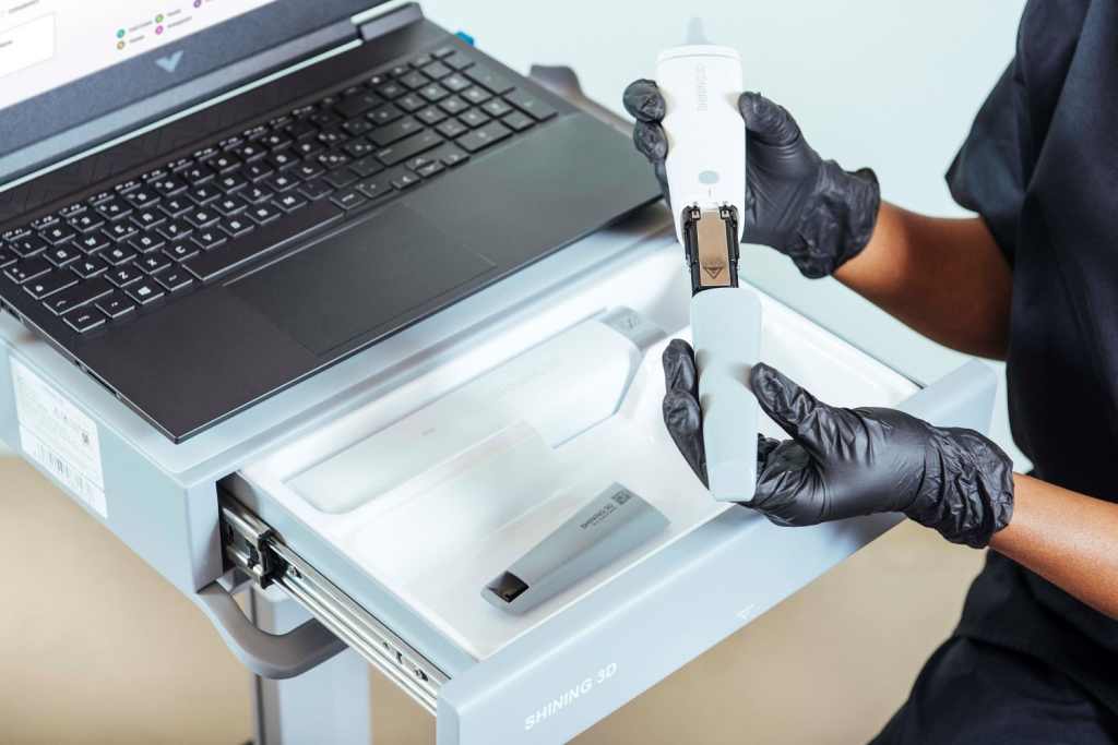
Fig 1: Two Tip sizes accommodate adults and children
As early as 2020, the dental program of Universidad de Cartagena and the PROMOUC research team collaborated with support from the Colombian Ministry of Innovation and Technology. Dr. Jadad and Dr. Sergio Tinoco planned a craniofacial reconstruction project for newborns, aimed at providing comprehensive care for patients with cleft lip and palate.
However, the project encountered significant challenges and risks during the process of taking dental impressions, particularly with regards to infants with cleft lip and palate (CLP). The materials and trays used for impressions impeded air circulation within the oral cavity, resulting in a heightened risk of choking or suffocation.
The use of the Aoralscan 3 will make this possible on Luna and Jesús. On January 1st, 2023, the collaborative efforts between Dr. Enrique Jadad Bechara, Dr. Sergio Tinoco, Dr. Sandra Cáceres, Dr Jorge Navarro officially began. They made their first attempt at treating Luna, a patient with unilateral cleft lip and palate, and Jesús, a patient with bilateral cleft lip and palate, using the Aoralscan 3 and its mini scan tip.
Within 24 hours of Luna and Jesús’ birth, Dr. Sandra Cáceres’ medical team initiated the care process and performed the first maxillary orthopaedic procedure, commonly known as the breastfeeding technique. Luna and Jesús’ mothers received education on how to help their babies latch onto the breast, which is beneficial for subsequent preoperative treatment using the obturator and enhances the newborns’ immune system during their growth and development.
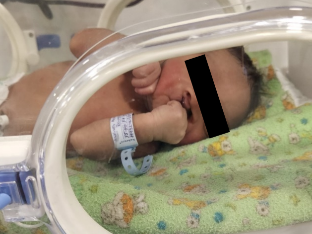
Fig 2: Jesús, male patient, 2 days old, with a diagnosis of complete bilateral cleft lip and palate
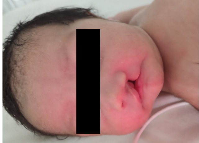
Fig 3: Luna, female patient, 2 days old, with a diagnosis of complete left unilateral cleft lip and palate
During the first step of the neonatal orthopedic phase, it is common to consider placing an obturator plate at the beginning of the treatment. This is done to preserve a normal dental arch and improve the baby’s feeding ability, preparing them for future surgeries. When Luna was 25 days old and Jesús was 23 days old, Dr. Enrique Jadad Bechara performed digital oral scans on them. Based on this data, custom-made obturator plates were created with the aim of improving the alignment of the back and front segments of the jaw, reducing the width of the cleft, and promoting breastfeeding.
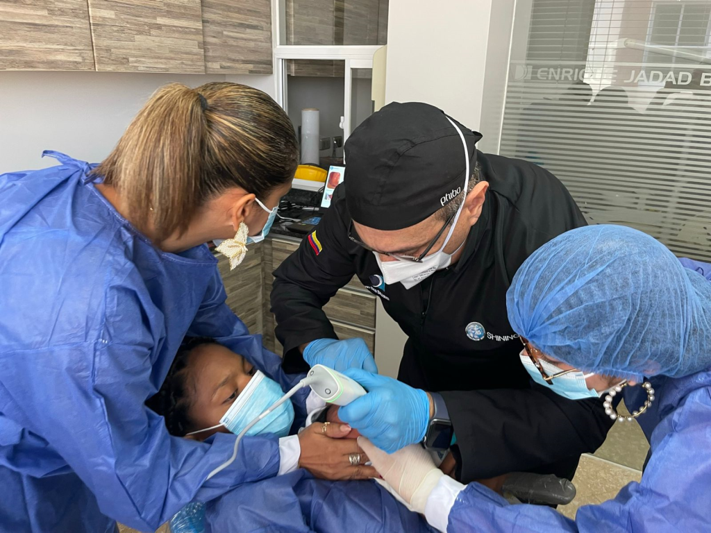
Fig 4: Dr Enrique Jadad performing intraoral scan with Aoralscan 3 and its mini scan tip for babies. The scanning depth of the Aoralscan 3 exceeded Dr. Jadad’s expectations.
In traditional physical impressions, meticulous control of the impression material properties and process is required to minimize the risk of newborn choking. However, digital intraoral scanners with their compact scan tips and rapid data acquisition enable precise intraoral data collection in a short amount of time. This method is not only fast and safe but also produces high-quality data.
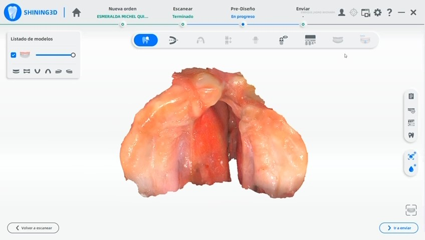
Fig 5: The intraoral data scanned by Aoralscan 3
The intraoral scan data was sent to Dr. Sergio Tinoco and his STLab laboratory. With the assistance of digital orthodontic software, they designed and manufactured obturator plates by predicting the movement of the baby’s bone blocks. This approach minimizes the separation of bone structure and reduces the risk of subsequent surgical treatments. These obturator plates were created entirely in a digital format, then 3D printed into physical objects using special resin materials and fitted to Luna and Jesús’s mouth.
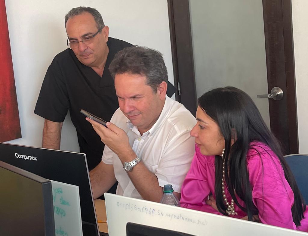
Fig 6: Dr. Sergio Tinoco and his team is analysis these two cases using professional orthodontic software.
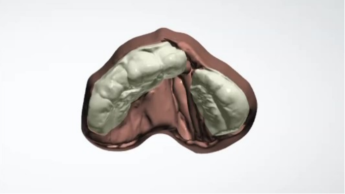
Fig 7: The simulation of bond movement in the software.
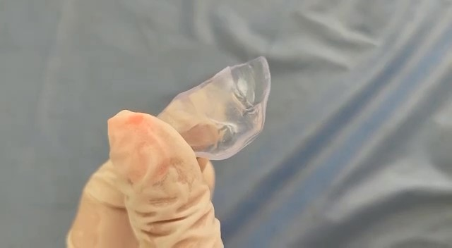
Fig 8: The printed obturator plates
These obturator plates not only help young patients improve their breastfeeding but also apply directed pressure and selective maxillary expansion, aiding in the reshaping of the maxillary bone, soft tissue, posterior alveolar segments, and anterior alveolar segments. Such a treatment process was unimaginable in the past.
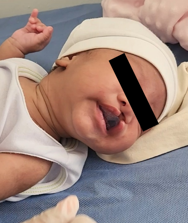
Fig 9: Luna is adapting the first phase obturator plates
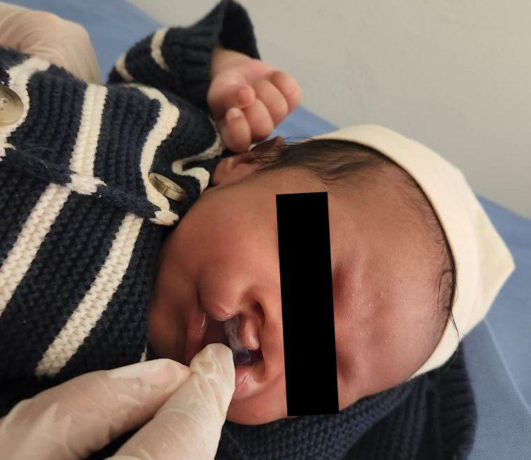
Fig 10: Jesús is adapting the first phase obturator plates
With the support of these obturator plates, these young patients were able to start breastfeeding immediately, ensuring sufficient nutrition. Luna, who had a unilateral cleft lip and palate, was able to latch onto her mother’s breast as soon as the obturator plate was installed. Just 8 days later, she reached the minimum weight required for the next surgical procedure. Jesús, who had a bilateral cleft lip and palate, took a few weeks to reach the minimum weight necessary for the surgery.
Afterwards, the surgical team at Harvard Plastic Surgery and the Unisinu Cartagena team led by Dr. Sandra Caceres performed surgery on Luna and Jesús, who were around 2 months old, to close the clefts in their upper lips.

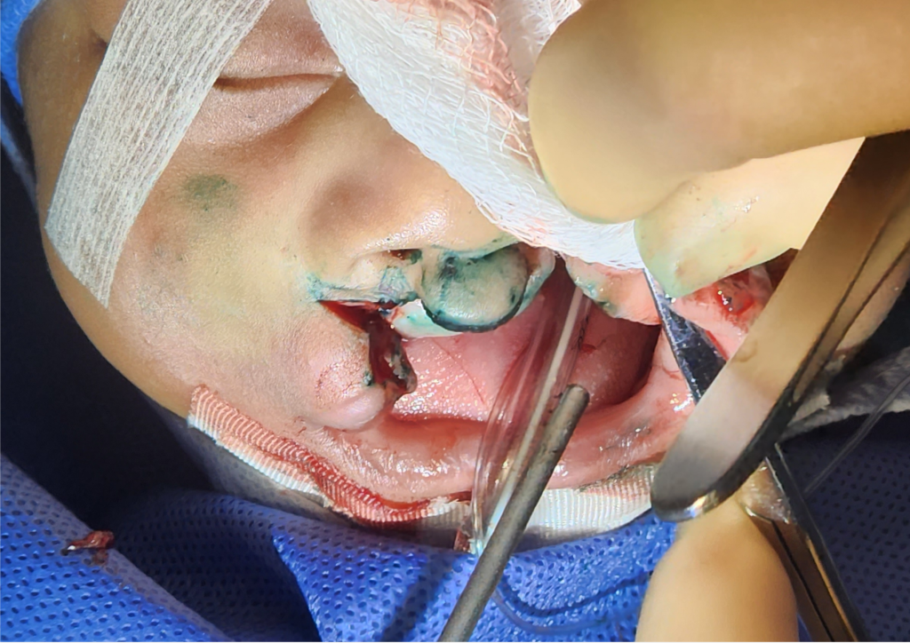
Fig 11,12: The surgical process to close the cleft in the upper lip for Luna and Jesús done by Harvard medical team
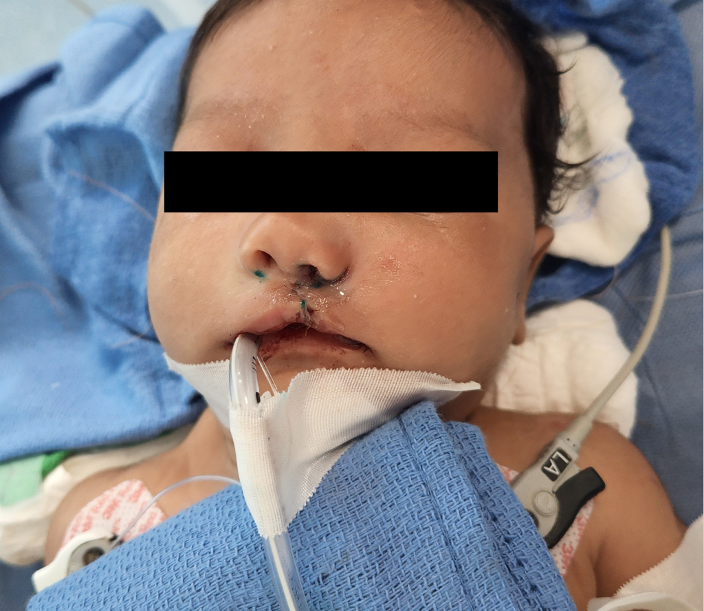
Fig 13: Luna after surgery.

Fig 14: Jesús after surgery.
After the surgery, Luna and Jesús will continue to wear obturator plates. By designing and using new obturator plates at different stages, they will help repair the cleft alveolar ridge, apply directed pressure, and guide the fractured maxillary bone to move towards the center, reducing the gap.
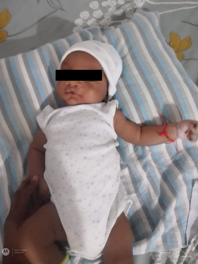
Fig 15: Jesús, good recovery after surgery to close the upper lip.
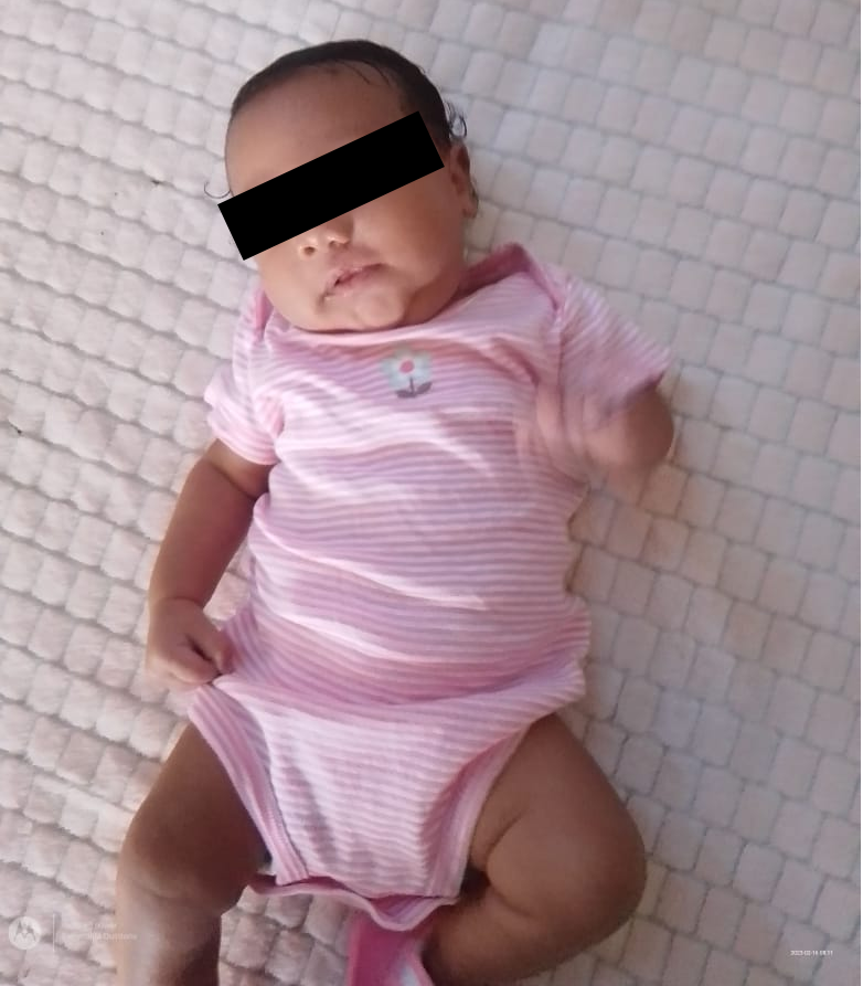
Fig 16: Luna, good recovery after surgery to close the upper lip.
Several months later, when Luna and Jesús were 5 months old, they underwent a second intraoral scan at Dr. Jadad’s clinic to analyze the progress made by the obturator plates in adjusting the alveolar ridge shape since the surgery. During this process, the medical team conducted individual assessments on Luna and Jesús, evaluating the suction and identifying the muscles of the intraoral and jaw system that needed strengthening to help both infants achieve the ideal weight for the second palatoplasty surgery. The initial proposal is to perform palatoplasty surgery when they are 1 year old in Jan. 2024.
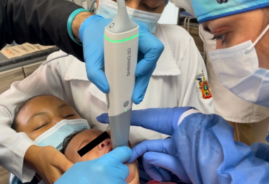
Fig 17: The second intraoral scan by Dr. Jadad to analyze the progress made by the obturator plates in adjusting the alveolar ridge shape since the surgery.
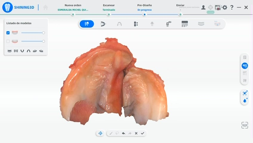
Fig 18: The Luna’s intraoral scan data in the second scan.
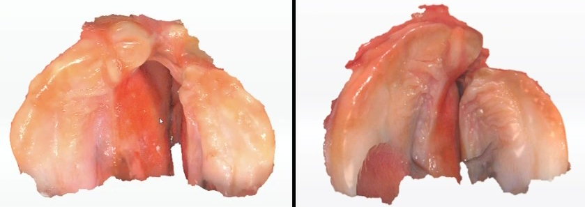
Fig 19: The comparison of Luna’s intraoral scan data before and after placing the obturator plates for about 4 months. (Left: first scan in Jan. 25th, 2023. / Right: second scan in Jun.28th, 2023. )
The application of digital technology, especially the use of intraoral scanners, along with a fully digital workflow based on printers and software, can reduce the separation of the maxilla caused by the defects present in Luna and Jesús at birth. This significantly shortens the recovery time required for cleft lip and palate patients. The surgical team from Harvard University predicts that the entire rehabilitation period will be reduced from 7 years to 2 years.
In this process, SHINING 3D’s Aoralscan 3 proves to be an outstanding and valuable tool, showcasing advanced technology, AI-based software, and a unique Mini scan tip that is unmatched in the global market.
About Author
The work team members are
- Dr. Sandra Cáceres, the research director at Universidad de Cartagena in Colombia.
- Dr. Sergio Tinoco, a specialist in orthodontics and maxillofacial surgery and the founder of ST LAB Digital Technologies.
- Jorge E Navarro B, Cirujano oral y Maxilofacial, Universidad Nacional Autónoma de México, Fundador y director científico Fundación Hagamos Sonreir.
- Dr. Enrique Jadad Bechara, Specialist in Oral Rehabilitation, researcher and lecturer with private practice in Barranquilla (Colombia), the KOL of SHINING 3D, along with the medical team from Harvard University.
It is a large and diverse group of professionals from various health and technology fields who have come together, leveraging digital technology to provide the best and most innovative comprehensive treatment for children with this type of morphological abnormality.