 ENG
ENG


Table of Contents
Since the launch of the Elite, we’ve been expanding the range of implant brands compatible with our IPG technology. While we’ve made great progress, there are still some implant MUAs that we’re unable to work with at this time.
Today’s case, provided by Dr. Dimitris Tasakos, is a great example of working with both compatible and non-compatible MUAs within the same arch. He demonstrates a method for combining the IPG workflow with the traditional implant workflow during the scanning process, expanding the Elite’s application for these kinds of patients.
Dr. Dimitris Tasakos is a DSD Master and ICOI Fellow, renowned for his active involvement in lectures and hands-on seminars on Digital Dentistry. His expertise spans Digital Impressions, Digitally Guided Implantology, and prosthetic restoration of implants, with a particular focus on direct loading protocols. Dr. Tasakos operates a dental clinic in Athens, where he offers comprehensive treatment using advanced digital technologies.
Case Description
A 61-year-old male patient had a 10-year-old metal-ceramic screw-retained implant prosthesis on the maxilla with multiple porcelain chipping areas that had been repaired many times in the past. His main concern was to replace it with a new implant-supported prosthesis.
Three years ago, he received six implants in the mandible and a zirconia full-contour prosthesis on them, and he desired the same type of prosthesis for the upper jaw. We know well that this type of prosthesis requires excellent accuracy, therefore, a very accurate and precise impression of the six upper implants was needed. To achieve outstanding accuracy, we decided to use SHINING3D’s Aoralscan Elite Intraoral Photogrammetry technology.
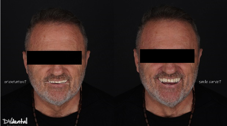
Additionally, due to the skewed smile line and unattractive appearance of the previous maxillary prosthesis, the patient expressed a desire for a more aesthetically pleasing design for the new restoration. To achieve this, we will use SHINING3D’s MetiSmile facial scanner to capture the patient’s facial data and use this data as a reference for smile design.
Treatment Process
1. Aoralscan Elite to capture all-on-six impression data
For occlusal reconstruction cases, precise data are the foundation of success. The intraoral scanner Aoralscan Elite’s IPG technology can provide sub-millimeter accuracy, ensuring the passive fit of the prosthesis, particularly in edentulous implant-supported restoration cases.
In this case, we encountered a problem: while the MUA of the four anterior implants were compatible with the IPG scan bodies, the two posterior implants were not compatible with the IPG scan bodies. Therefore, we will scan the two posterior implants using normal 3D reconstruction technology (in the soft tissue scan, placing two standard scan bodies on the MUA of the two posterior implants) and then scan the anterior implants with IPG technology (placing the IPG scan bodies on the MUA of the four anterior implants).
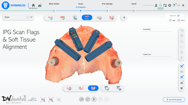
In the next stage, alignment between the IPG scan bodies and the soft tissue, along with the standard scan bodies, will be performed to obtain a digital model with six scan bodies of the Implants.
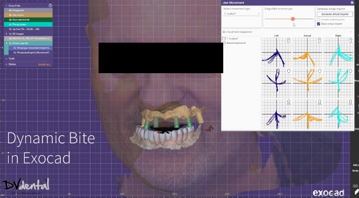
To achieve more precise occlusion, in the final step of the scanning process, Aoralscan Elite can help us capture the dynamic bite, which can be imported into the design software for subsequent dynamic occlusal design.

Fig 4: First step, scan the Pre-op scan to capture the old restoration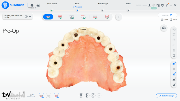
Fig 5: Second step, scan the lower jaw
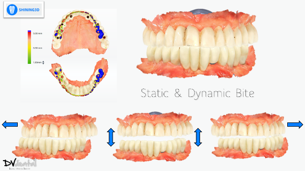
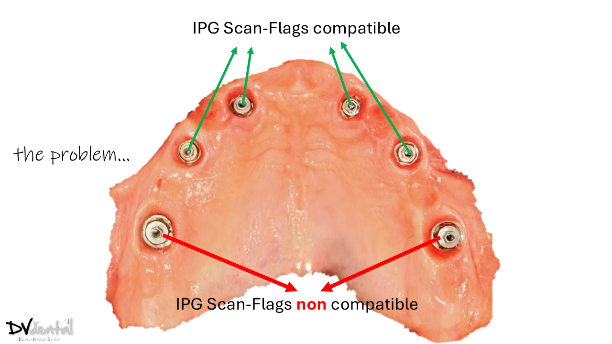
Fig 7: In the same arch, four MUAs are coded scanbody compatible, while the rest two MUAs are coded scanbody non compatible.

Fig 8, 9: Step four, extra scan match to capture the emergence file and align with the pre-op scan

Fig 10: Step five, scan the soft tissue and normal scan flags.
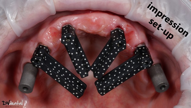
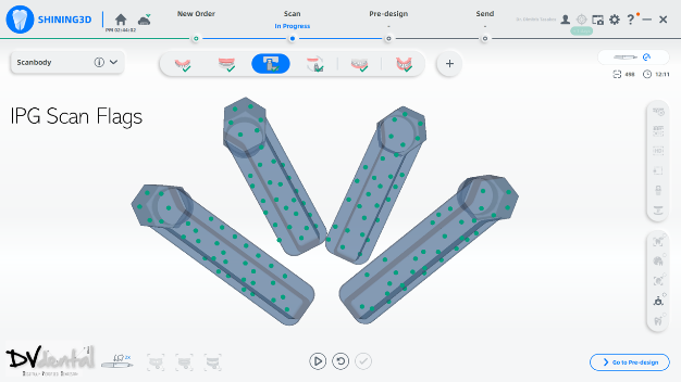
Fig 11,12: Step 6, install and scan the coded scanbody using photogrammetry technology
2. MetiSmile and acquisition of facial data
In aesthetic design, facial data can assist technicians in analyzing the patient’s facial symmetry, smile line, and the proportion of teeth to facial features, ensuring that the prosthesis harmonizes with the patient’s facial characteristics. Therefore, the application of facial scanners is crucial. The MetiSmile facial scanner from Shining 3D can capture the patient’s 3D facial data with high precision, including facial contours, lip morphology, nasolabial folds, and chin position. These data provide essential references for the aesthetic design of the prosthesis. Since both MetiSmile and Aoralscan Elite belong to the Shining 3D platform, the intraoral scan data and facial scan data can be directly matched during the facial scanning process.

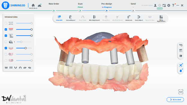
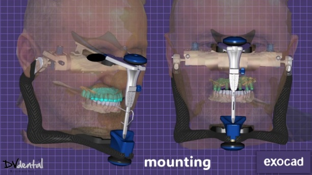
3. Design in exocad
After the capture process is done, we send everything to the lab. The lab mounts the data on the virtual articulator with the help of the face-scan, and designs a facially driven prosthesis for the upper jaw according to the patient’s needs. The integration of the dynamic bite within the CAD software allowed us to finalize our design functionally.

4. Try-in
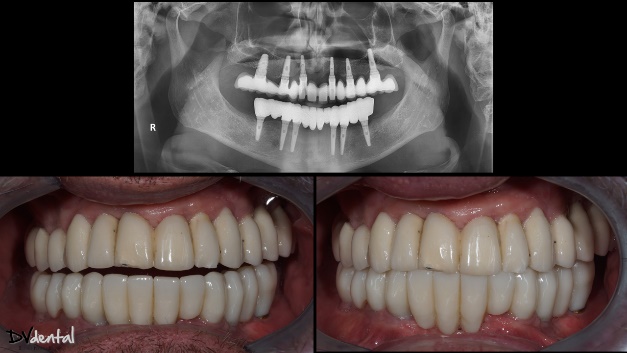
Before the permanent prosthesis, a prototype of it has been created for intraoral try-in. This is a good way to check the fit of our prosthesis clinically and radiographically while assessing the aesthetics simultaneously. After the successful try-in, the manufacturing of the permanent prosthesis (Zirconia full contour on Ti bases) will proceed. Finally, periapical X-rays show that the permanent prosthesis fits excellently, and the aesthetic outcome was exactly as planned, the patient is really happy about the result.

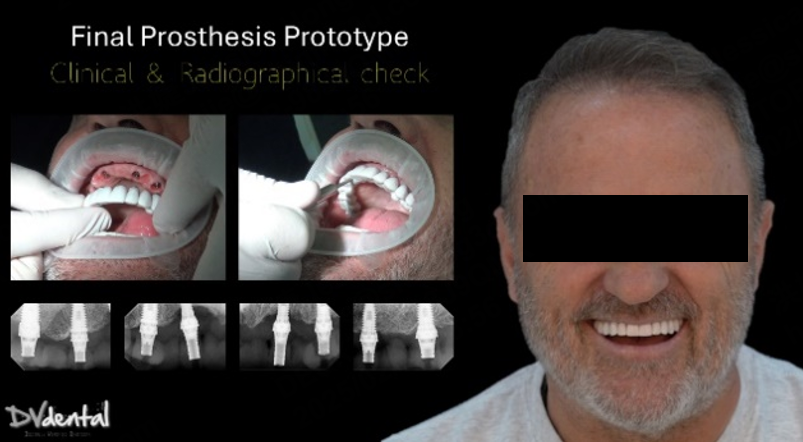
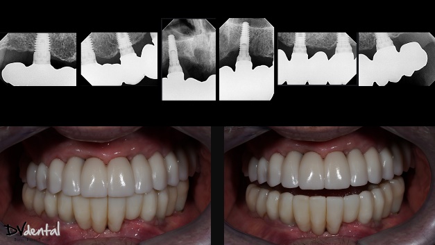
Fig 22: The intraoral photos after treatment.

The comments from Dr. Dimitris Tasakos:
The benefits of MetiSmile and Aoralscan Elite in all-on-X implant cases
The Elite intraoral scanner, combine with IPG technology, provided a highly accurate digital impression for this case, laying the foundation for the implant restoration design. The MetiSmile face scanner thoroughly captured the patient’s facial morphology, ensuring the restoration’s harmony in both function and aesthetics, making it an essential tool in the occlusal reconstruction process.
This case highlights the significant advantages of digital technology in full edentulous implant restoration. Specifically, the combined use of the intraoral scan with IPG technology and facial scanning offers a precise, efficient, and personalized solution for complex occlusal reconstruction cases.