 ENG
ENG


The advancement of material and digital technology makes it possible for patients to retain as much of their own natural teeth while having ultra-thin lithium disilicate veneers. Today’s case from Dr. Luis Estuardo Pacheco, an experienced dentist who specializes in aesthetics and implantology. Within this case study, you will see how he delivered ultra-thin veneers to a young patient with minimally invasive teeth preparation.
Case Profile and Treatment Plan
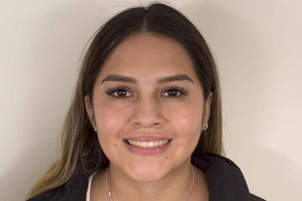
Fig 1: The initial situation before treatment.
The patient, a 23 young woman who was not satisfied with the apperance of her yellow teeth. Upon further observation, she had suffered from abrasion in both the upper jaw and lower jaw, which lead to hypersensitivity when chewing. The case’s goal was to recover the original beauty and the normal function of her teeth. The initial plan was to place ultra-thin veneers in the upper jaw using digital smile design and to have the lower jaw repaired later when she was able to afford it.
Treatment Process – Intraoral Scan & Face Scan
The first step is minimally invasive tooth preparation, followed by data collection. Capturing all the necessary information is vital for an accurate digital smile design. In this case, the dentist obtained the intraoral scan data using the Aoralscan 3. This aids the dentist in getting rid of the physical impression, saving time, material cost, and it is more accurate. In order to successfully capture the 3D facial data, the dentist scanned the patient’s face . The scanner is fast, accurate, and creates a high resolution 3D facial scan in full color.
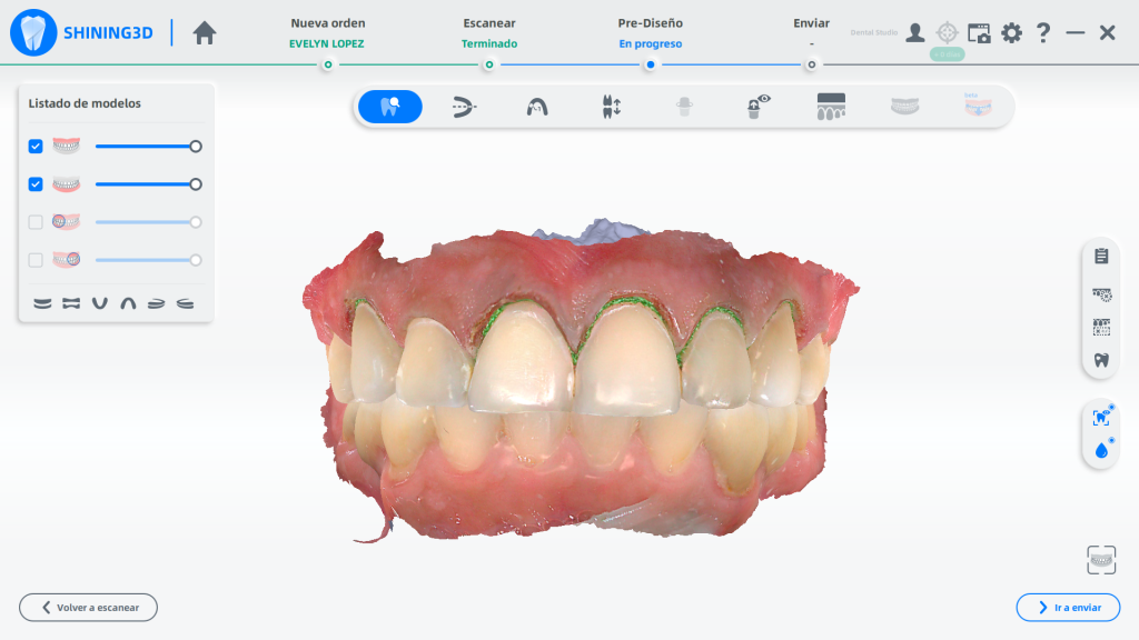
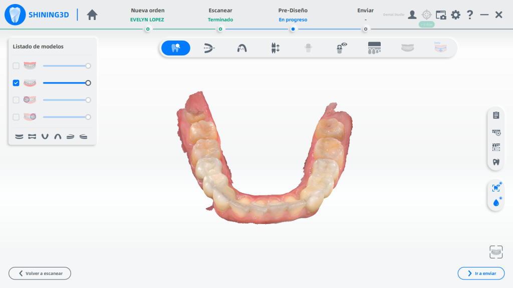
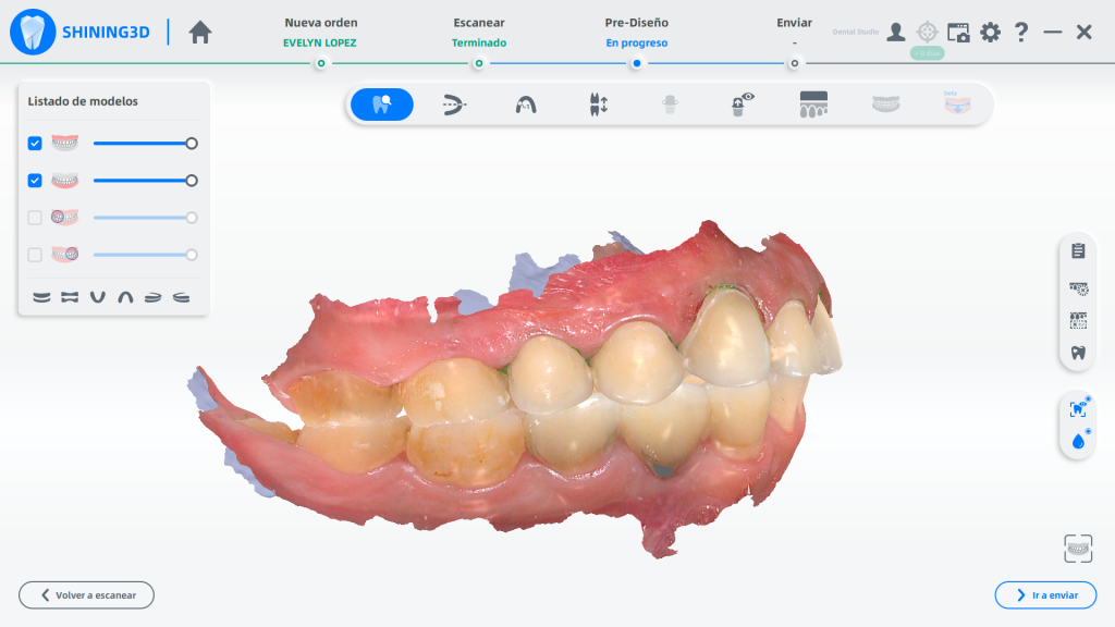
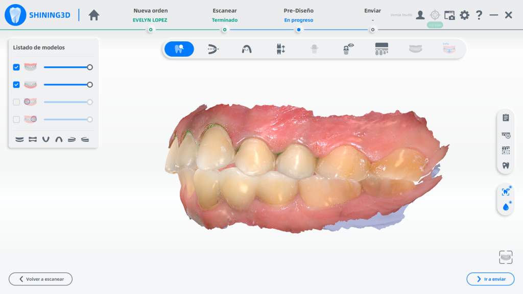
Fig 2,3,4,5: The intraoral data is captured by Aoralscan 3, the figures above show the front view, the occlusion view, and the left and right side.
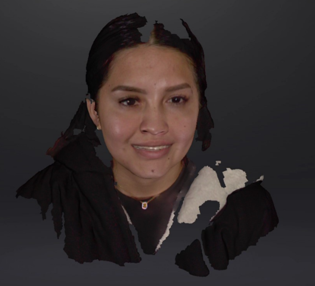
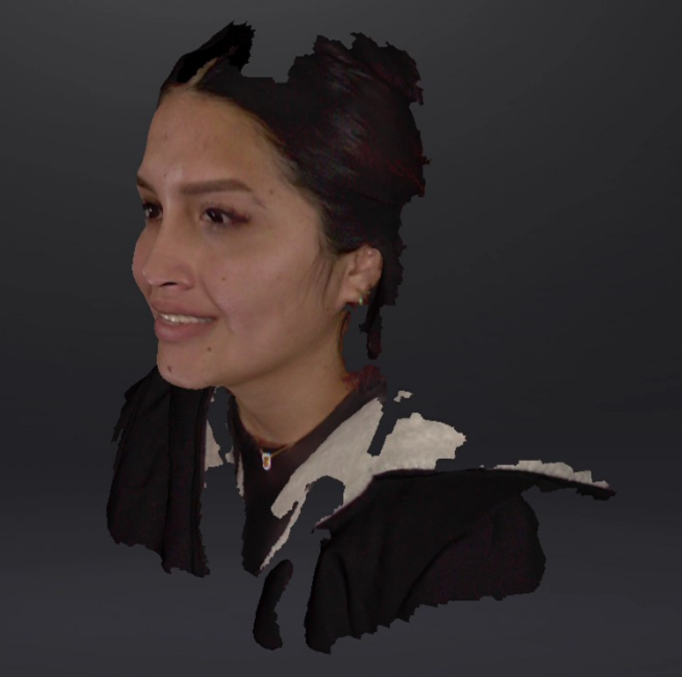
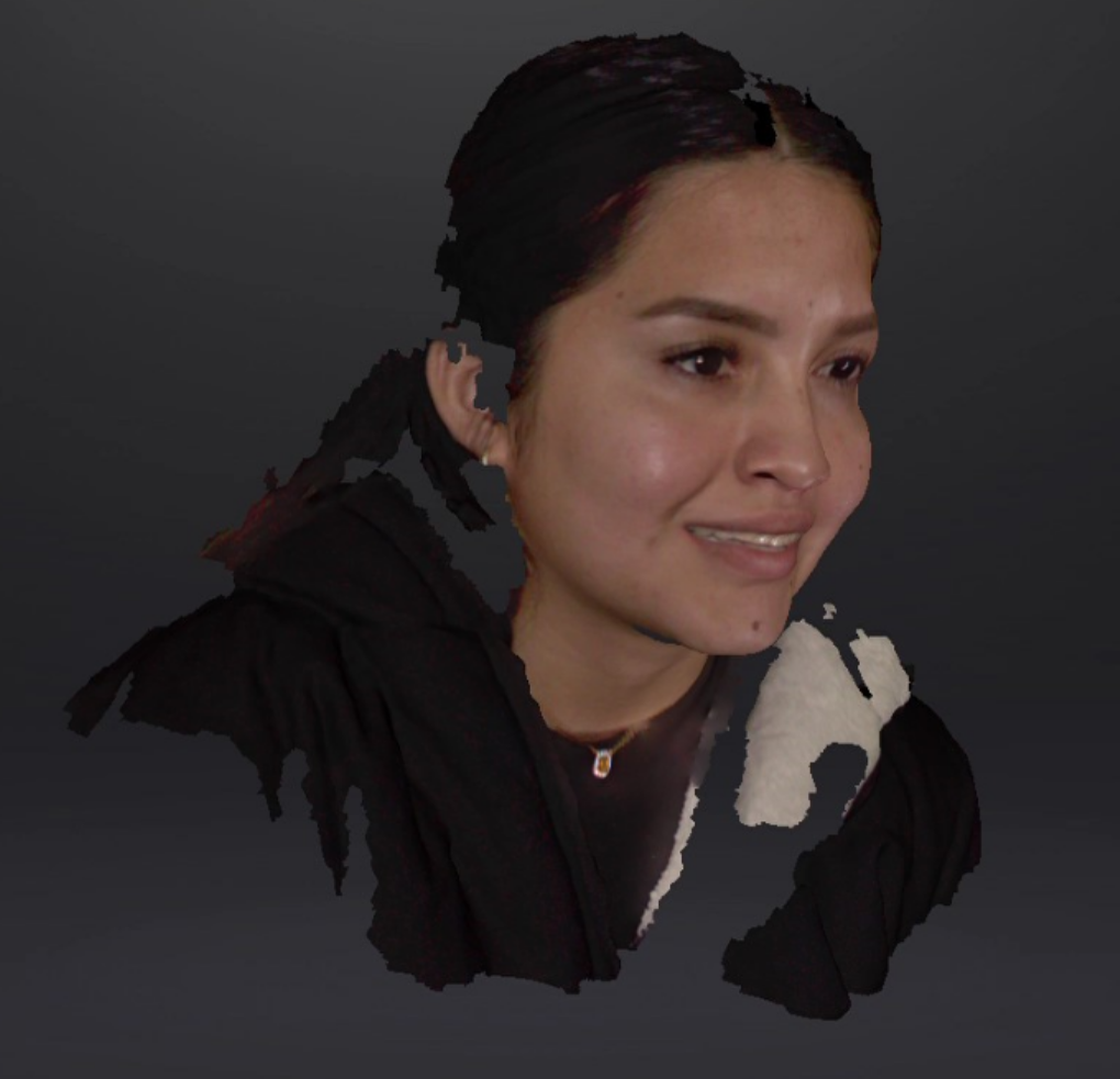
Fig 6,7,8: The face scan data captured by MetiSmile.
Treatment Process — Digital Smile Design
To have #15~#25 restored in ultra-thin veneers, the dentist had the digital intraoral scan model and face scan model imported into exocad for professional design.
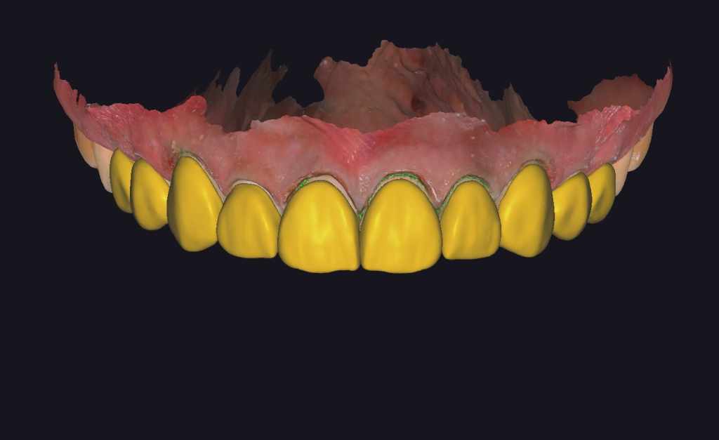
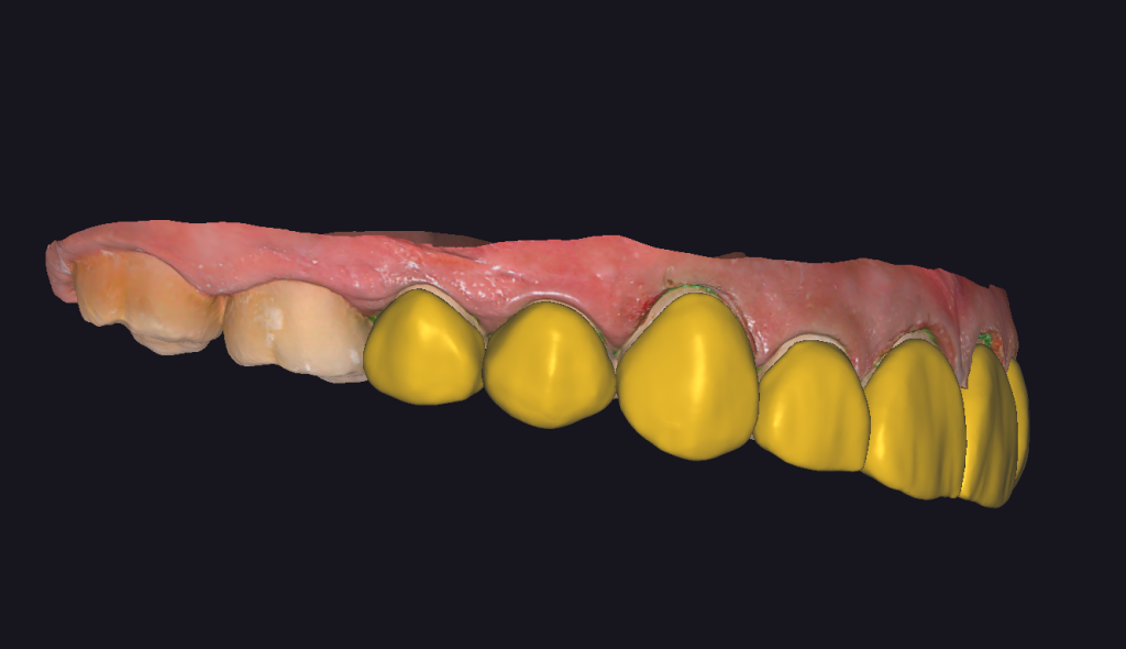
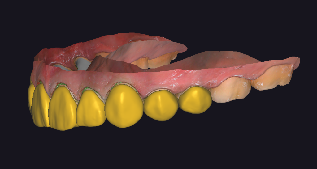
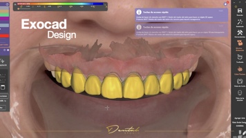
Fig 9,10,11,12:The design process in exocad.
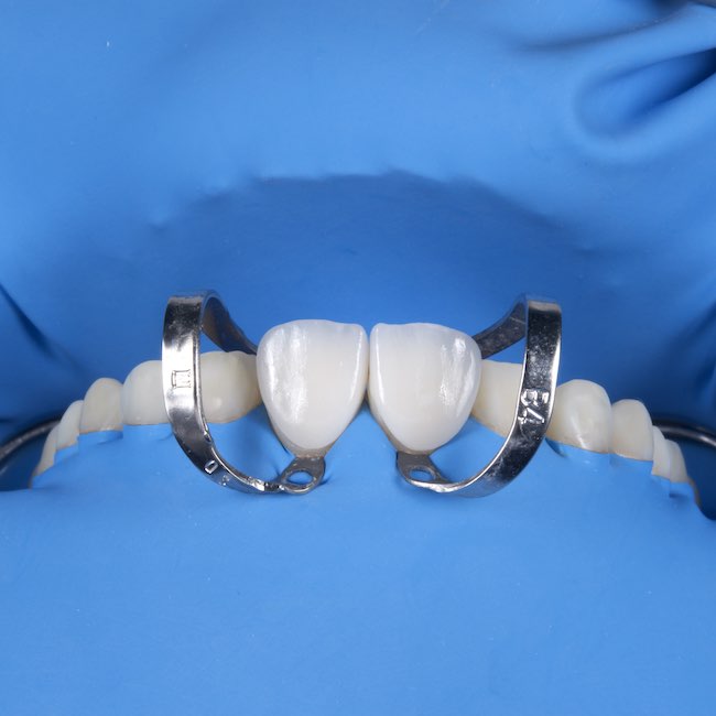
Treatment Process – Veneer Delivery
The veneers appear very bright and white, as the patient preferred to have a shining outlook. After a standard cementing process, the patient was very satisfied with the final restoration result.
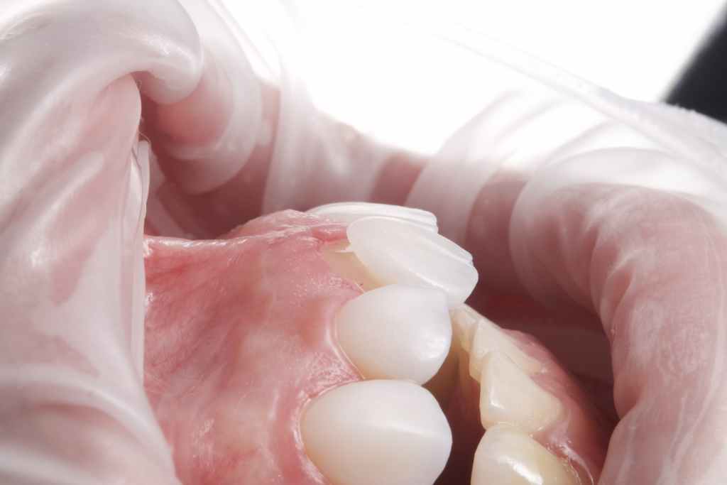
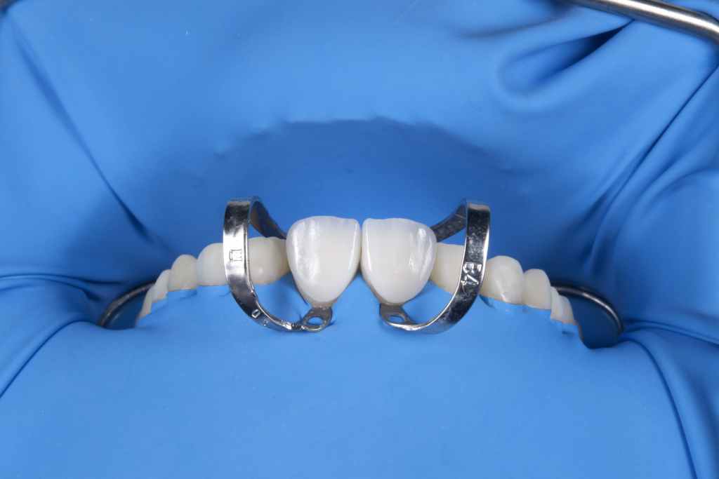
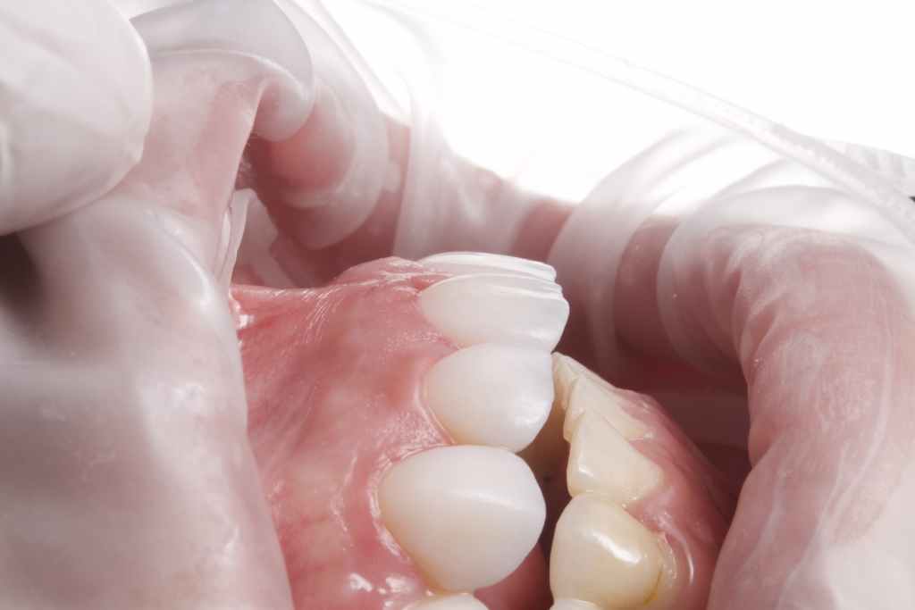
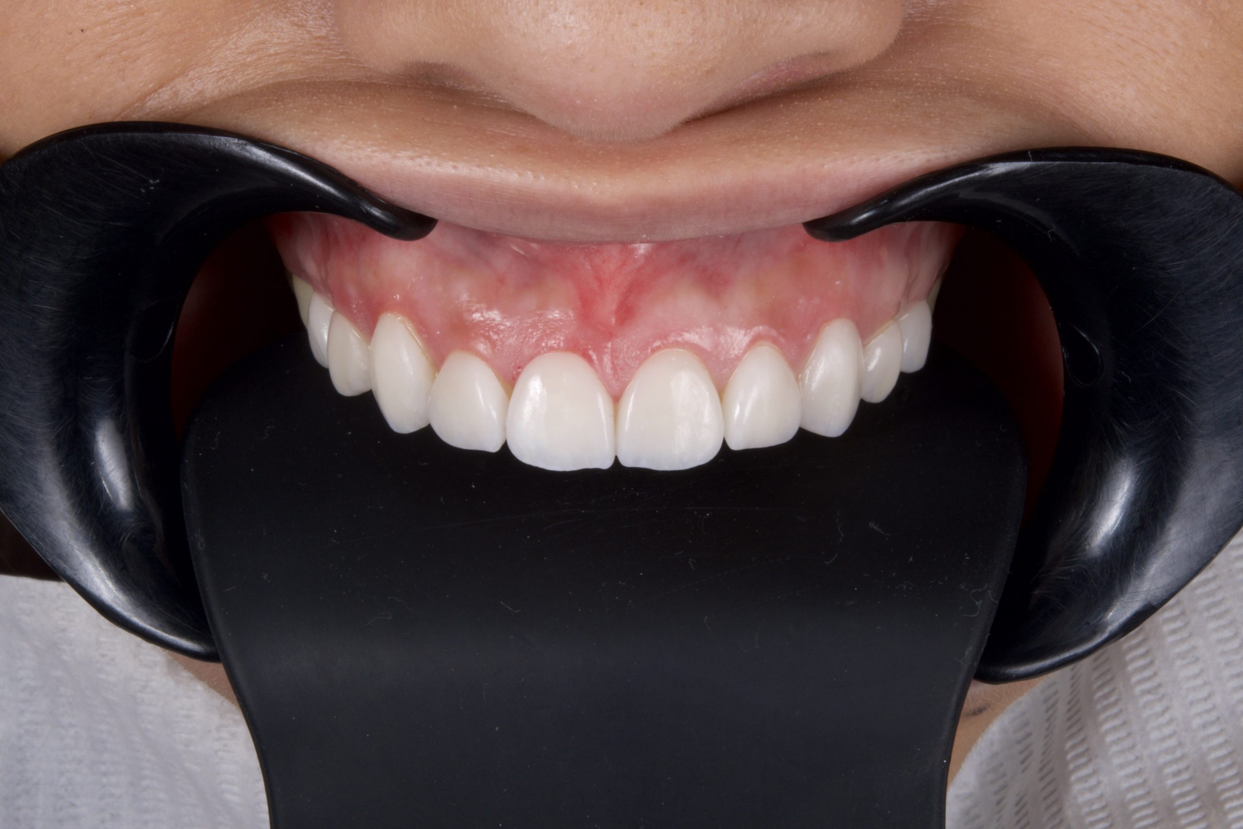
Fig 13,14,15,16:The process to fix the ultra-thin veneers to the patient.
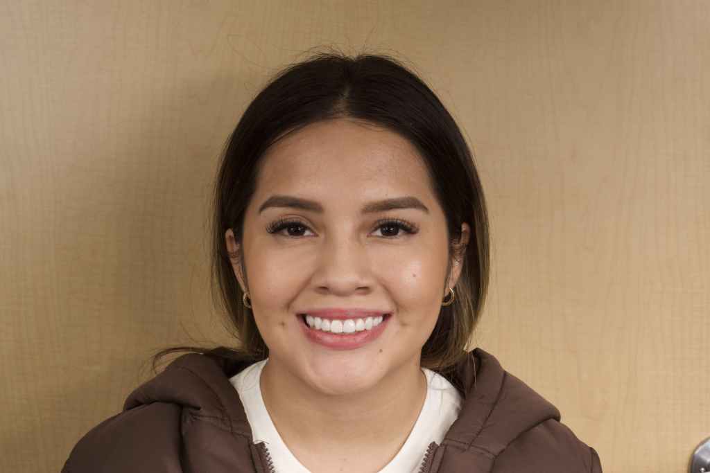
Fig 17: After treatment
Case summary
A fully digital workflow is currently the fastest and most accurate way to provide predictable and satisfying results for both dentists and patients in aesthetic cases. The advantage of intraoral and facial scanning includes time-management, increased accuracy, and reducing human error, especially in challenging cases. In this case, an accurate design is made based on intraoral scan data combined with face scan data, and advanced milling techniques, which help make the manufacturing possible.
About Author
- Dr. Luis Estuardo Pacheco
- DDS., MSc.
- CEO at Dental Studio, Guatemala.
- Senior CAD/CAM consultant, Scientific Team Lab(STL), 3D Labs CAD/CAM Laboratories, Guatemala.
- Senior consultant in oral and maxillofacial radiology at 3D Xray, Guatemala.
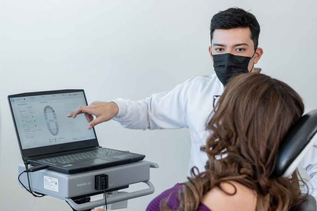
Fig 18: Dr. Luis Estuardo Pacheco