



Case provided by Dr. Ayah Taha Abdallah
Introduction
Modern implant dentistry is increasingly guided by a "start with the end in mind" philosophy, where prosthetically driven planning dictates the entire surgical and restorative workflow. The integration of digital diagnostic setups, dual-scan protocols, and CBCT imaging establishes a highly predictable blueprint for full-arch rehabilitations. To seamlessly translate this virtual design into clinical reality, precise data acquisition is paramount. Intraoral photogrammetry (IPG) serves as a critical bridge in this process. By eliminating the dimensional distortions inherent in conventional long-span impressions, IPG guarantees the absolute passive fit of the final prosthesis, ensuring that the initial aesthetic and functional goals are flawlessly achieved.
Clinical Situation
A 64-year-old male patient , presented to the clinic with severe dental debilitation. His chief complaint was explicitly stated as, "I can neither eat well nor smile".
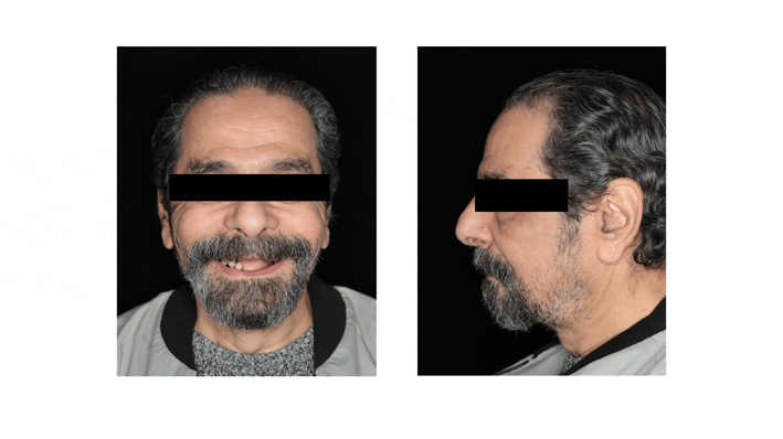
Intraoral examination revealed that only a few compromised teeth remained. Guided by a prosthetically driven approach, all remaining teeth were planned for extraction to facilitate a full-arch rehabilitation. The patient experienced significant functional impairment alongside considerable aesthetic concerns regarding his smile.
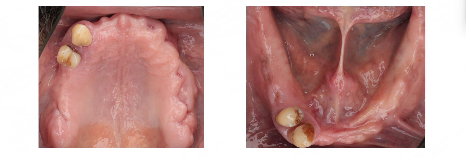
Diagnostics and Virtual Planning
The planning phase began with establishing jaw relation records and the appropriate vertical dimension of occlusion, followed by a conventional diagnostic teeth setup. These dentures were subsequently duplicated to function as radiographic stents, embedded with radiopaque markers.
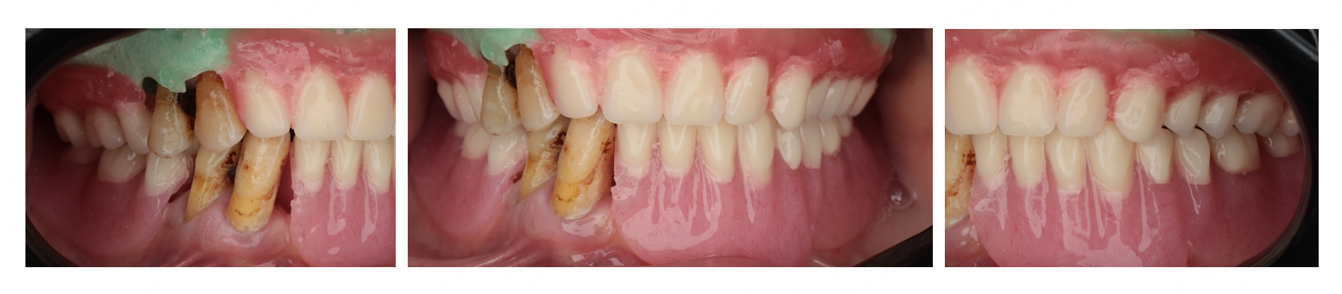
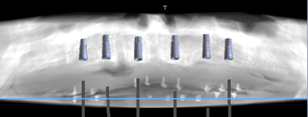
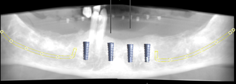
Digital superimposition of the patient's intraoral scans, the diagnostic denture, and the CBCT data was performed. This allowed the clinician to align the proposed teeth perfectly with the underlying bone structure.
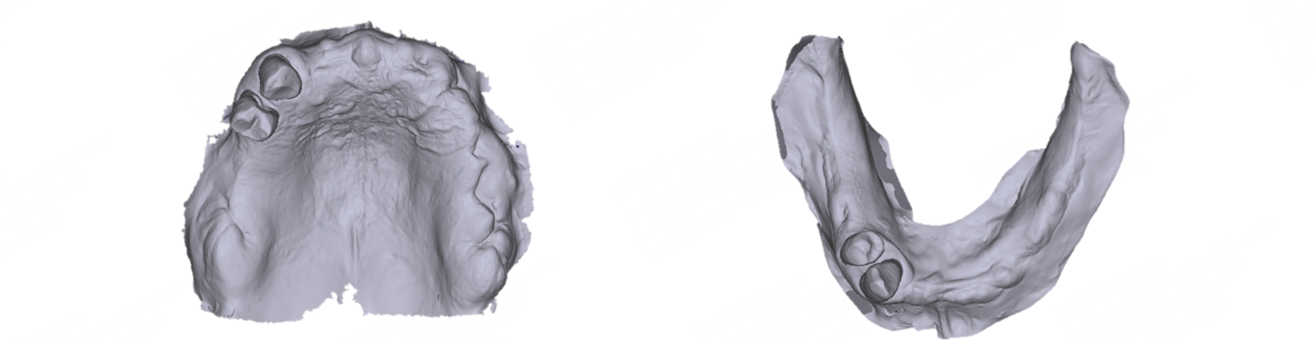
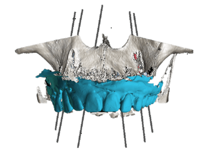
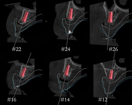
This achieved a high superimposition accuracy of 0.004. Based on this prosthetically driven setup, virtual implant planning was finalized to place four implants in the mandible and six implants in the maxilla. Specifically for the upper arch, implants were precisely planned for sites #12, #14, #16, #22, #24, and #26, utilizing 4x11.5mm dimensions.
Surgical Procedure
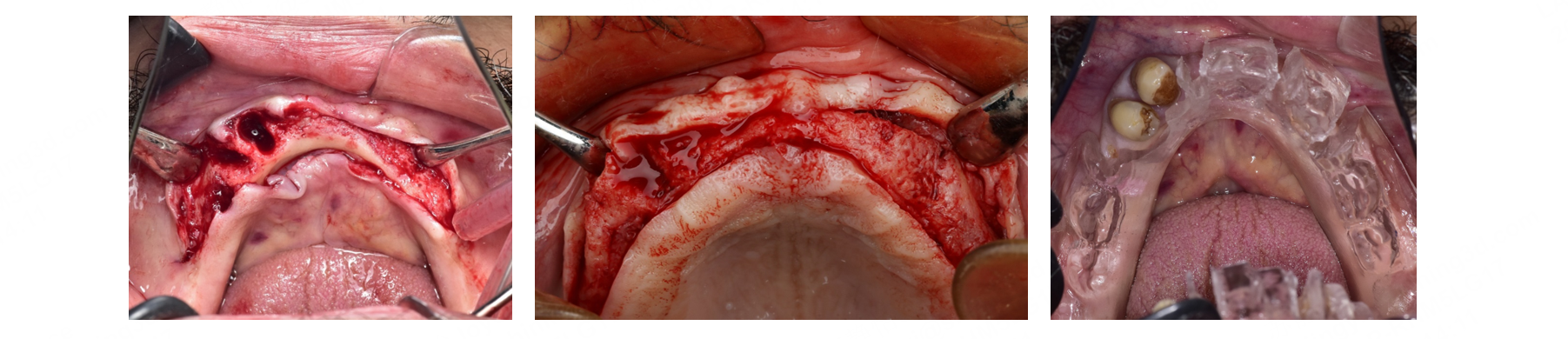
Due to patient-related factors, the surgical intervention was staged; the mandibular implants were placed first, and the maxillary surgery followed at a later date. For both arches, after the extraction of the remaining compromised teeth and full-thickness flap reflection, the duplicated radiographic stents were utilized intraoperatively to accurately mark the planned points of insertion.
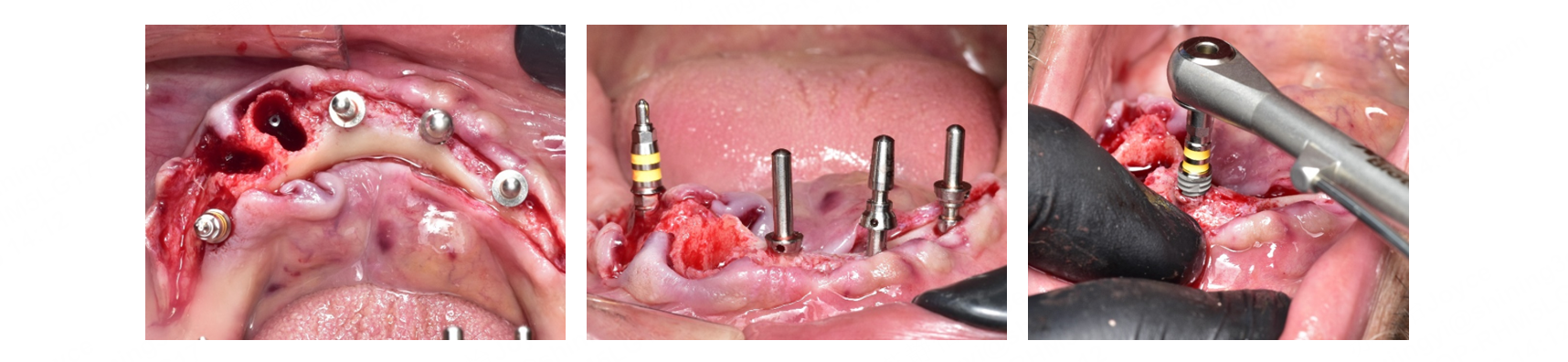
Following site marking, osteotomies were prepared, and parallel tools were placed to visually verify implant angulation. The implants were then successfully placed via a free-hand surgical approach.
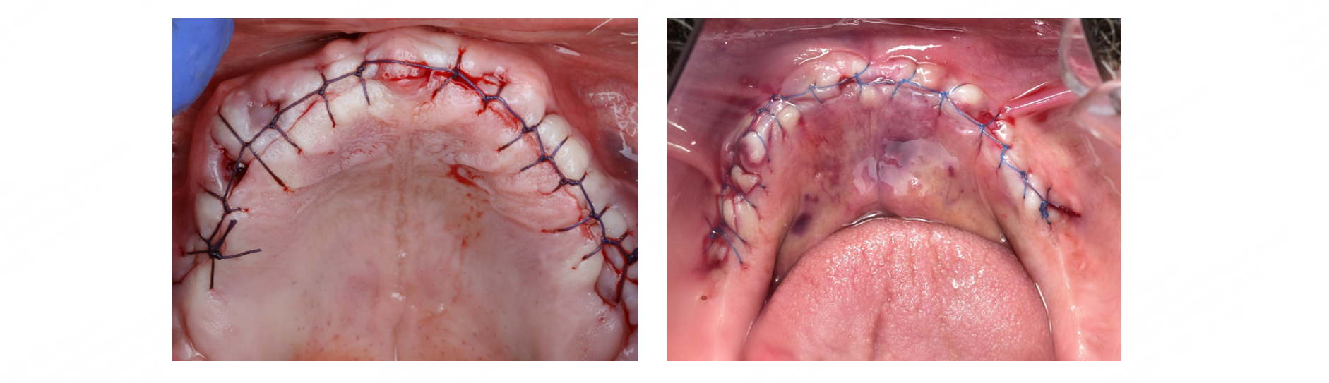
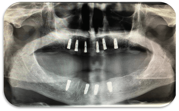
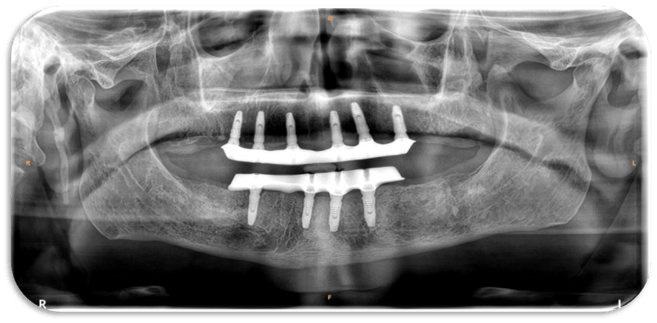
The surgical sites were immediately closed with sutures. A postoperative panoramic radiograph confirmed the successful insertion of the upper arch alongside the already osseointegrated lower implants.
Healing Phase and Prosthetic Release
Following successful osseointegration of the implants, second-stage surgery was conducted to place Multi-Unit Abutments (MUAs).
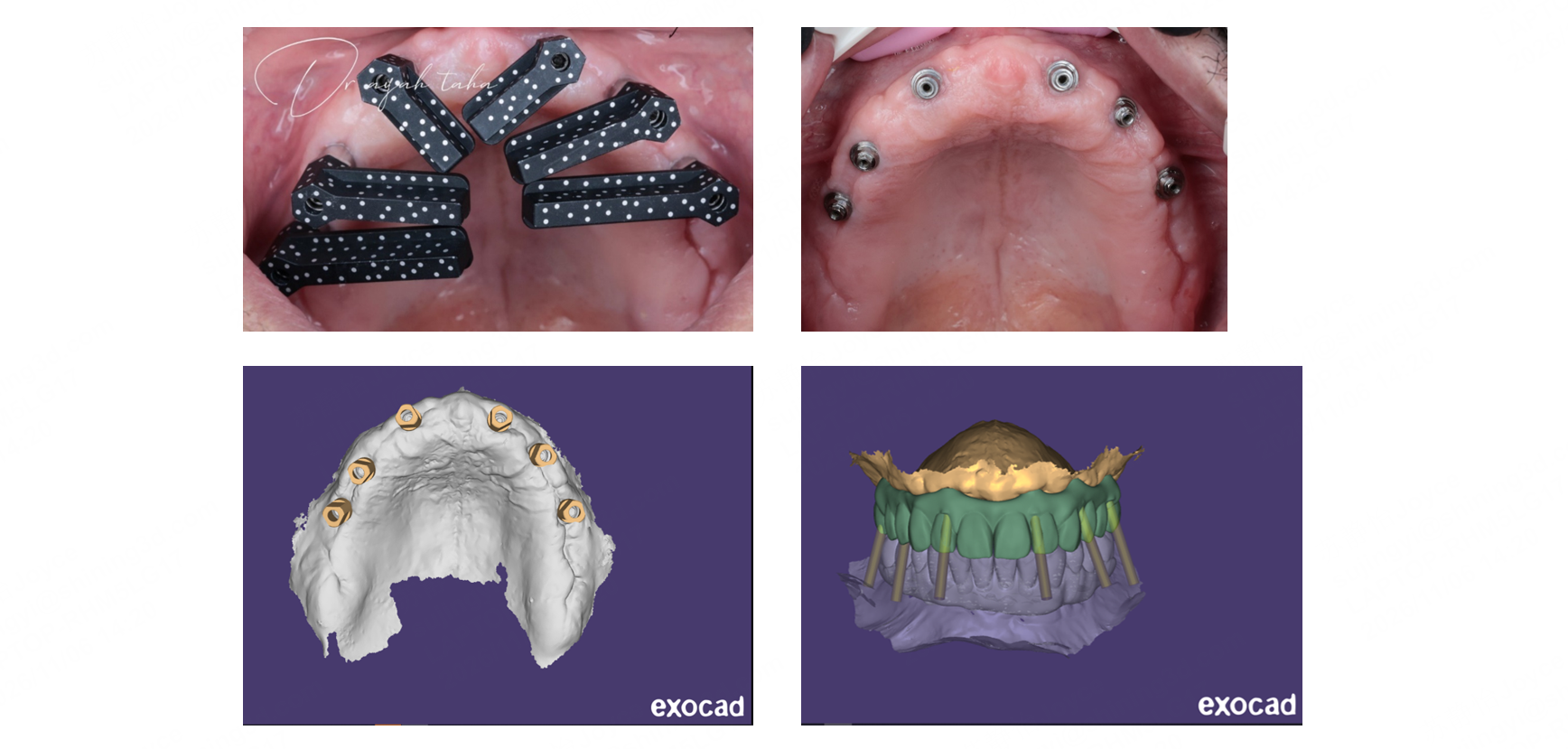
For the complex six-implant maxillary arch, Shining 3D intraoral photogrammetry (IPG)was utilized. The use of IPG scan bodies significantly increased the accuracy of the scanning data capture and made the clinical workflow substantially easier and faster. This highly precise dataset was transferred to exocad for the final design.
A milled PMMA try-in was fabricated and seated to confirm the absolute passive fit of the framework.
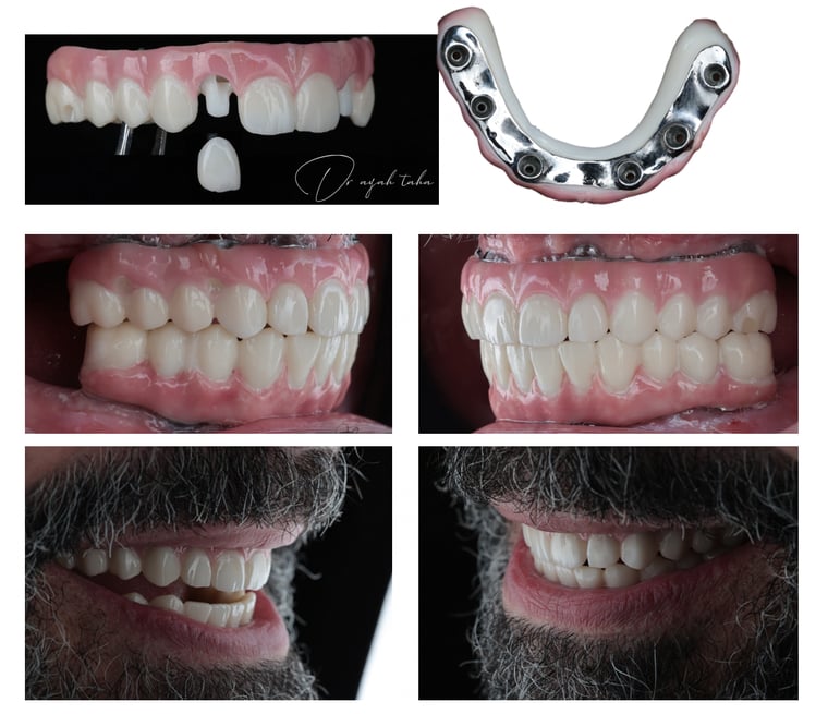
Finally, a high-aesthetics Zirconia over bar (bar split) prosthesis was permanently inserted, immediately restoring the patient's masticatory function and facial aesthetics.
Conclusion
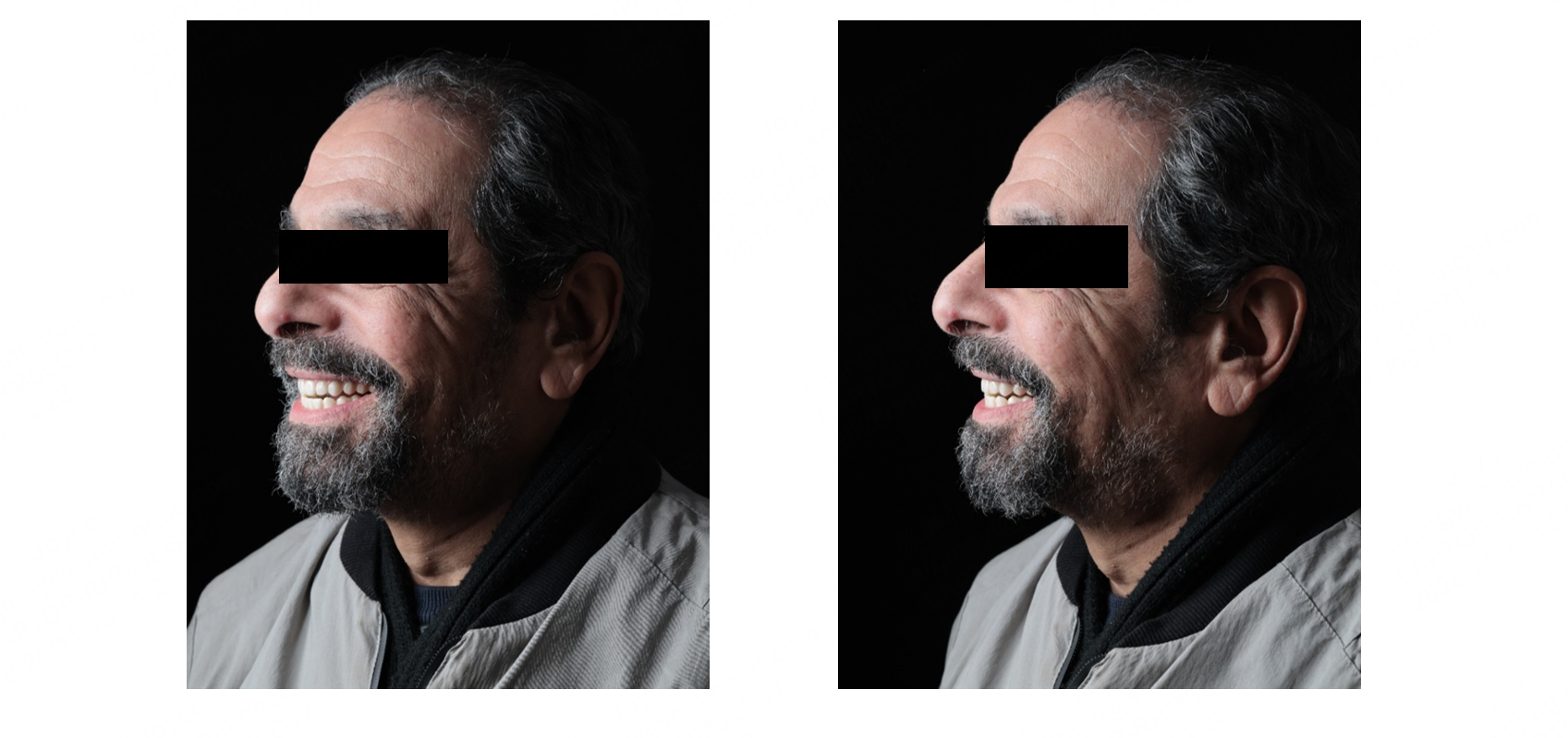
This case strongly highlights the clinical advantages of incorporating advanced data acquisition into full-arch rehabilitations. The strategic combination of CBCT imaging, dual-scan diagnostics, and staged surgical execution provided a safe, functional foundation. Furthermore, upgrading from conventional splinted scan bodies to intraoral photogrammetry (IPG) during the prosthetic phase minimized the cumulative errors associated with cross-arch scanning. This highly accurate digital workflow ensured the perfect passive fit of the final zirconia over bar framework, successfully restoring the patient's confidence, smile, and quality of life.
Share this on Social Media:


