 JPN
JPN


デジタルインプラント技術の開発に伴い、口腔内スキャンと手術ガイドの適用がますます人気になっている。インプラント手術では、口腔内データをもとに、患者様の口腔内の状態に合わせた外科手術ガイドを印刷することで、手術の精度と効率を高めることができる。
患者情報
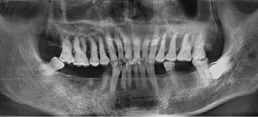
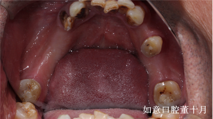
患者は両側の下後歯が4年前から欠損しており、CBCTは、右下4番歯の根管に高密度の光線遮断物質、根端の低密度影、唇側骨壁の完全な骨吸収が判明した。右下5番歯の歯槽堤の上部には直径1mmの残存根があり、右下5番と7番歯、左下4番と6番歯の歯槽堤の幅は適度であった。
インプラント手術のワークフロー
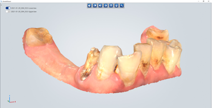
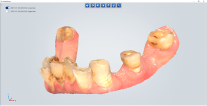
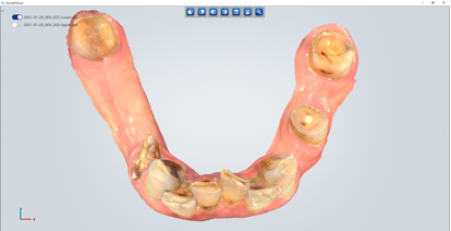
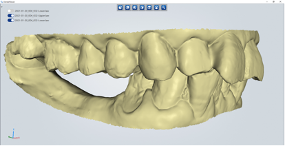
デジタル印象を取得する
引き続きの手術ガイドの設計のために、Aoralscanで口腔内データを取得する。
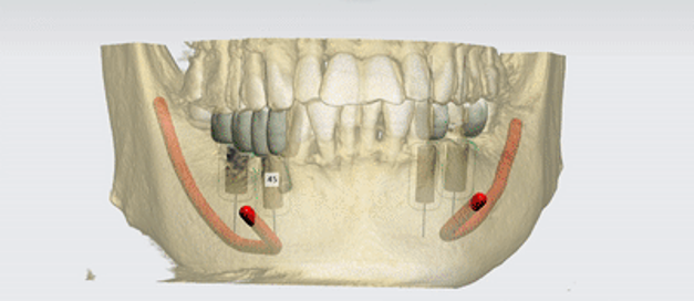
治療計画
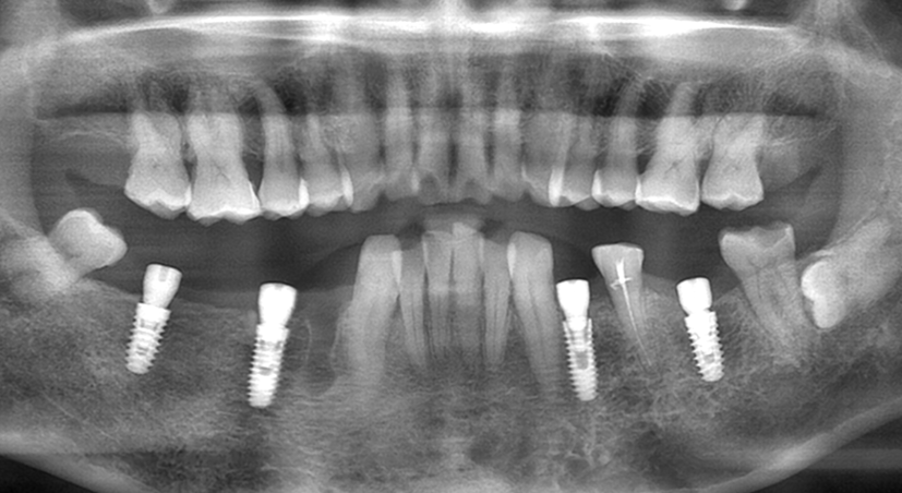
実際の骨量に応じて、歯の位置左下6番歯、右下7番歯に4.8 * 10のインプラントを配置する。十分な垂直的骨量があるため、4 * 12&4.3 * 12のインプラントを歯の位置左下4番と右下5番歯に配置する。手術後、延期されたインプラントの埋入のために右下4番歯(残存根)が抜き取られる。
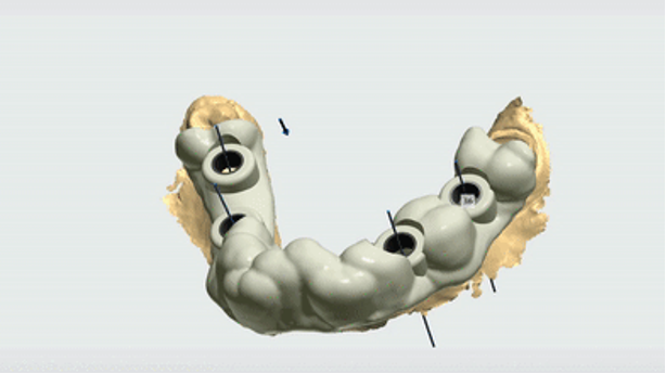
手術ガイドを設計する
ソフトウェアによって生成された外科手術用ガイドは分割できなくて、そしてガイドのスパンが大きいために、位置決めが難しいである。したがって、ガイドは、外科医が手術中にガイドを簡単に取り外せるには、3番目のソフトウェアによって2つの部分に分割する。
手術ガイドを印刷する
歯科用3DプリンターAccuFabで手術ガイドを印刷する

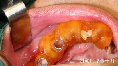
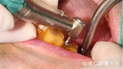
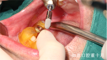
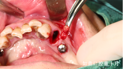
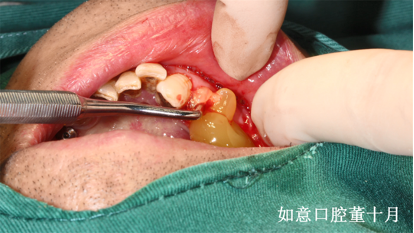
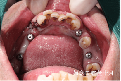
インプラント手術
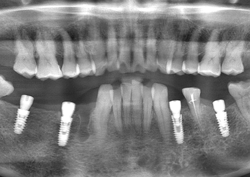
術後CT検査
Due to the irregular shape of the alveolar ridge, the absorption of labial bone on #34, insufficient width of alveolar ridge and uneven bone density at the implant bed area, in addition, the patient has a certain degree of mouth opening limitation, manual operation may lead to poor orientation and affect the final restoration. With the help of surgical guide, the orientation and depth can be precisely controlled and, more importantly, it’s minimally invasive and it can also reduce the operation time and the fear of patient in the process of surgery.